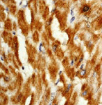
ab76152 at 1/50 dilution staining PDK2 in human heart muscle by Immunohistochemistry, Paraffin-embedded tissue.
![Anti-PDK2 antibody [EP1947Y] (ab76152) at 1/1000 dilution + SH-SY-5Y cell lysate at 10 µgSecondarygoat anti-rabbit HRP at 1/1000 dilutiondeveloped using the ECL technique](http://www.bioprodhub.com/system/product_images/ab_products/2/sub_4/9332_ab76152wb.gif)
Anti-PDK2 antibody [EP1947Y] (ab76152) at 1/1000 dilution + SH-SY-5Y cell lysate at 10 µgSecondarygoat anti-rabbit HRP at 1/1000 dilutiondeveloped using the ECL technique
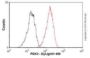
Overlay histogram showing HepG2 cells stained with ab76152 (red line). The cells were fixed with 80% methanol (5 min) and then permeabilized with 0.1% PBS-Tween for 20 min. The cells were then incubated in 1x PBS / 10% normal goat serum / 0.3M glycine to block non-specific protein-protein interactions. The cells were then incubated with the antibody (ab76152, 1/100 dilution) for 30 min at 22ºC. The secondary antibody used was DyLight® 488 goat anti-rabbit IgG (H+L) (ab96899) at 1/500 dilution for 30 min at 22ºC. Isotype control antibody (black line) was rabbit IgG (monoclonal) (1µg/1x106 cells) used under the same conditions. Acquisition of >5,000 events was performed. This antibody gave a positive signal in HepG2 cells fixed with 4% paraformaldehyde (10 min)/permeabilized with 0.1% PBS-Tween for 20 min used under the same conditions.