Anti-PDIA6 antibody
| Name | Anti-PDIA6 antibody |
|---|---|
| Supplier | Abcam |
| Catalog | ab135594 |
| Prices | $384.00 |
| Sizes | 400 µl |
| Host | Rabbit |
| Clonality | Polyclonal |
| Isotype | IgG |
| Applications | IHC-P WB FC |
| Species Reactivities | Mouse, Human |
| Antigen | Synthetic peptide conjugated to KLH, corresponding to a region within internal sequence amino acids 151-180 of Human PDIA6 |
| Description | Rabbit Polyclonal |
| Gene | PDIA6 |
| Conjugate | Unconjugated |
| Supplier Page | Shop |
Product images
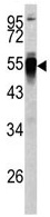 Anti-PDIA6 antibody (ab135594) at 1/50 dilution + Y79 cell line lysate at 35 µg
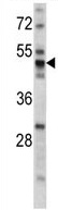 Anti-PDIA6 antibody (ab135594) at 1/50 dilution + Mouse stomach tissue lysate at 35 µg
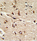 Immunohistochemical analysis of Formalin-fixed, Paraffin-embedded Human brain tissue labelling PDIA6 with ab135594 at 1/50 dilution followed by peroxidase-conjugated secondary antibody and DAB staining.
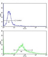 Flow cytometric analysis of HeLa cells labelling PDIA6 with ab135594 at 1/50 dilution (bottom histogram) compared to a negative control cell (top histogram). FITC conjugated Goat anti Rabbit secondary antibodies were used for the analysis.