
Overlay histogram showing HeLa cells stained with ab137132 (red line). The cells were fixed with 80% methanol (5 min) and then permeabilized with 0.1% PBS-Tween for 20 min. The cells were then incubated in 1x PBS / 10% normal goat serum / 0.3M glycine to block non-specific protein-protein interactions followed by the antibody (ab137132, 1/100 dilution) for 30 min at 22°C. The secondary antibody used was Alexa Fluor® 488 goat anti-rabbit IgG (H&L) (ab150077) at 1/2000 dilution for 30 min at 22°C. Isotype control antibody (black line) was rabbit IgG (monoclonal) (1μg/1x106 cells) used under the same conditions. Unlabelled sample (blue line) was also used as a control. Acquisition of >5,000 events were collected using a 20mW Argon ion laser (488nm) and 525/30 bandpass filter.
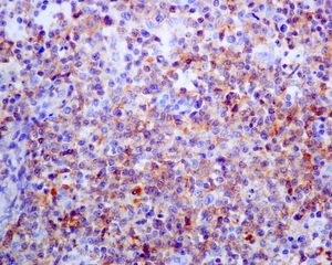
Immunohistochemical analysis of paraffin embedded Human T-cell lymphoma tissue labelling PD1 using ab137132 at a dilution of 1/250.
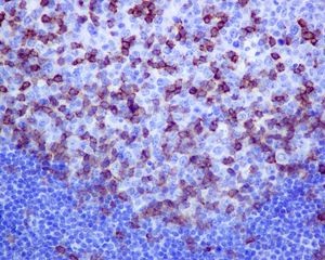
Immunohistochemical analysis of paraffin embedded Human tonsil tissue labelling PD1 using ab137132 at a dilution of 1/250.
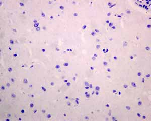
Immunohistochemical analysis using ab137132 showing negative staining in Normal brain tissue.
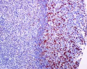
Immunohistochemical analysis using ab137132 showing positive staining in Normal tonsil tissue.

Immunohistochemical analysis using ab137132 showing negative staining in Skeletal muscle tissue.