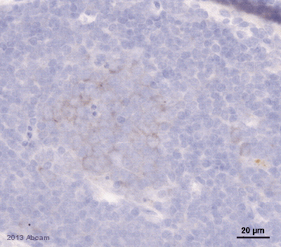
ab117420 staining PD1 in Mouse spleen tissue sections by Immunohistochemistry (IHC-P - paraformaldehyde-fixed, paraffin-embedded sections). Tissue was fixed with formaldehyde and blocked with 2.5% serum for 1 hour at 25°C; antigen retrieval was by heat mediation in a citrate buffer pH 6. Samples were incubated with primary antibody (1/100 in blocking buffer) for 12 hours at 4°C. An undiluted HRP-conjugated Rabbit anti-goat IgG polyclonal was used as the secondary antibody.See Abreview

ab117420, at 5 ug/ml, staining PD1 in Formalin-fixed, Paraffin-embedded Human Small Intestine (Peyer's Patch) tissue by Immunohistochemistry followed by biotinylated secondary antibody, alkaline phosphatase-streptavidin and chromogen.
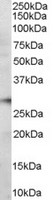
Anti-PD1 antibody (ab117420) at 0.6 µg/ml + Human Bone Marrow lysate in RIPA buffer at 35 µgdeveloped using the ECL technique