Anti-PCNA antibody
| Name | Anti-PCNA antibody |
|---|---|
| Supplier | Abcam |
| Catalog | ab2426 |
| Prices | $400.00 |
| Sizes | 1 ml |
| Host | Rabbit |
| Clonality | Polyclonal |
| Isotype | IgG |
| Applications | IHC-F IP WB IHC-P ICC/IF ICC/IF |
| Species Reactivities | Mouse, Rat, Hamster, Dog, Human, Zebrafish, Marmoset, Fish |
| Antigen | Synthetic peptide: DMGHLKYYLAPKIEDEEGS , corresponding to C terminal amino acids 243-261 of Human PCNA |
| Description | Rabbit Polyclonal |
| Gene | PCNA |
| Conjugate | Unconjugated |
| Supplier Page | Shop |
Product images
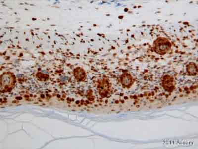
 ab2426 staining PCNA in Mouse 14.5 dpc tissue sections by Immunohistochemistry (IHC-P - paraformaldehyde-fixed, paraffin-embedded sections). Tissue was fixed with formaldehyde, permeabilized with TBS-T and blocked with 1% BSA + 1% FBS in TBS for 2 hours at room temperature; antigen retrieval was by heat mediation in Tris buffer, pH 9. Samples were incubated with primary antibody (1/1000 in blocking buffer) for 16 hours at 4°C. An undiluted HRP-conjugated Goat anti-rabbit IgG polyclonal was used as the secondary antibody.See Abreview
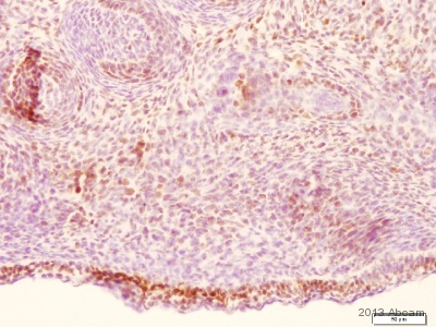
 ab2426 staining PCNA in murine skin tissue by Immunohistochemistry (Formalin/PFA-fixed paraffin-embedded sections). Tissue was fixed with formaldehyde and a heat mediated antigen retrieval step was performed using TRIS EDTA pH 8.2. Samples were then incubated with the primary antibody at a 1/2000 dilution for 1 hour at 37°C. An undiluted HRP-conjugated rabbit polyclonal was used as secondary antibody.See Abreview
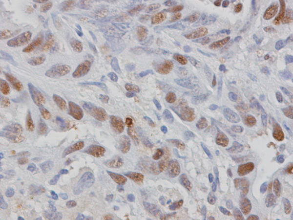 ab2426 staining PCNA in rat tumor tissue sections by Immunohistochemistry (IHC-P - paraformaldehyde-fixed, paraffin-embedded sections). Tissue was fixed with formaldehyde and blocked with 3% serum for 30 minutes at 20°C; antigen retrieval was by heat mediation in a citrate buffer, pH 6.0. Samples were incubated with primary antibody (1/500 in PBS) for 12 hours at 4°C. A HRP-conjugated goat anti-rabbit IgG polyclonal (1/200) was used as the secondary antibody.See Abreview
 ab2426 was used for immunohistochemistry, at a 1:500 dilution in hamster neural tissue, identified with a polyclonal secondary and DAB detection kit. Review by Kevin Bath submitted 18 June 2004
 ab2426 at 1/200 dilution staining rat skeletal muscle satellite stem cells by ICC/IF.Cultured rat skeletal muscle satellite stem cells (SKMB) were 2% paraformaldehyde fixed for 15 minutes and then permealized with Triton-X100 prior to incubation with ab2426 overnight at 4°C. A donkey anti-rabbit Alexa-Fluor ® 488 (ab150073) antibody was used as the secondary.The image shows DAPI (blue-nuclear stain, upper left panel), PCNA (Green, upper right panel, showing nuclear localization in actively dividing cells), same SKMB cells -DIC (phase) image (lower left panel) and superimpose fluorescence image (lower right panel).See Abreview
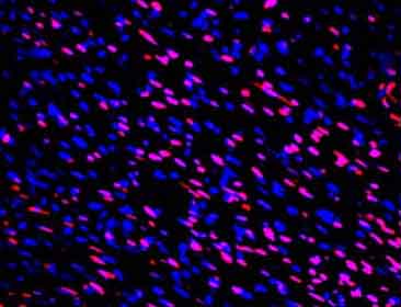 ab2426 staining PCNA in murine epithelial tissue by Immunohistochemistry (Formalin/PFA-fixed paraffin-embedded sections).Wound tissue sections (0.5 µm) were cut using a microtome and collected on slides. Sections were then de-waxed in xylene and rehydrated by successive immersion in descending concentrations of alcohol. The sections were then subjected for immunofluorescence staining. Briefly, tissue sections were blocked by incubated with 5% donkey serum (ab7475) for 1 hour and washed with phosphate-buffered saline (PBS). Sections were then incubated with ab2426 at a 1/500 for 1 hour at room temperature under humidified conditions. After primary antibody incubation, sections were washed with PBS and incubated with appropriate fluorescent secondary antibodies for 1 hour at room temperature. Sections were then washed with PBS, mounted with mounting medium containing DAPI.
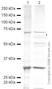 All lanes : Anti-PCNA antibody (ab2426) at 1/200 dilutionLane 1 : HeLa (Human epithelial carcinoma cell line) Nuclear LysateLane 2 : HeLa (Human epithelial carcinoma cell line) Whole Cell LysateLysates/proteins at 20 µg per lane.SecondaryIRDye 680 conjugated Goat anti-rabbit IgG (H&L) at 1/15000 dilution
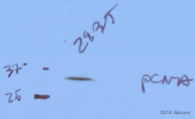 Anti-PCNA antibody (ab2426) at 1/1000 dilution + 293T whole cell lysate at 70 µgSecondaryHRP-conjugated goat anti-rabbit IgG polyclonal at 1/2000 dilutiondeveloped using the ECL techniquePerformed under reducing conditions.
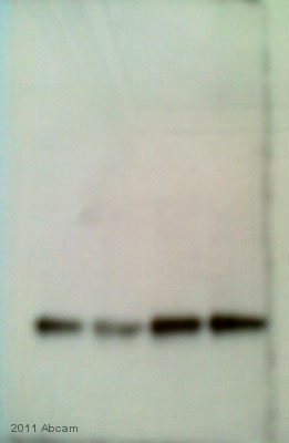 All lanes : Anti-PCNA antibody (ab2426) at 1/300 dilutionLane 1 : Whole tissue lysate prepared from mouse heartLane 2 : Whole tissue lysate prepared from mouse heartLane 3 : Whole tissue lysate prepared from mouse heartLane 4 : Whole tissue lysate prepared from mouse heartLysates/proteins at 50 µg per lane.SecondaryHRP-conjugated pig anti-rabbit polyclonal at 1/3000 dilutiondeveloped using the ECL technique
Product References
IL-15 deficient tax mice reveal a role for IL-1alpha in tumor immunity. - IL-15 deficient tax mice reveal a role for IL-1alpha in tumor immunity.
Rauch DA, Harding JC, Ratner L. PLoS One. 2014 Jan 8;9(1):e85028.
Quantification of healthy and atretic germ cells and follicles in the developing - Quantification of healthy and atretic germ cells and follicles in the developing
Inserra PI, Leopardo NP, Willis MA, Freysselinard AL, Vitullo AD. Reproduction. 2013 Dec 20;147(2):199-209.
Chemopreventive evaluation of a Schiff base derived copper (II) complex against - Chemopreventive evaluation of a Schiff base derived copper (II) complex against
Hajrezaie M, Hassandarvish P, Moghadamtousi SZ, Gwaram NS, Golbabapour S, Najihussien A, Almagrami AA, Zahedifard M, Rouhollahi E, Karimian H, Fani S, Kamalidehghan B, Majid NA, Ali HM, Abdulla MA. PLoS One. 2014 Mar 11;9(3):e91246.
Olive oil and omega-3 polyunsaturated fatty acids suppress intestinal polyp - Olive oil and omega-3 polyunsaturated fatty acids suppress intestinal polyp
Barone M, Notarnicola M, Caruso MG, Scavo MP, Viggiani MT, Tutino V, Polimeno L, Pesetti B, Di Leo A, Francavilla A. Carcinogenesis. 2014 Jul;35(7):1613-9.
Emodin suppresses hyperglycemia-induced proliferation and fibronectin expression - Emodin suppresses hyperglycemia-induced proliferation and fibronectin expression
Gao J, Wang F, Wang W, Su Z, Guo C, Cao S. PLoS One. 2014 Apr 1;9(4):e93588.
Poldip2 knockout results in perinatal lethality, reduced cellular growth and - Poldip2 knockout results in perinatal lethality, reduced cellular growth and
Brown DI, Lassegue B, Lee M, Zafari R, Long JS, Saavedra HI, Griendling KK. PLoS One. 2014 May 5;9(5):e96657.
Role of microRNAs in resveratrol-mediated mitigation of colitis-associated - Role of microRNAs in resveratrol-mediated mitigation of colitis-associated
Altamemi I, Murphy EA, Catroppo JF, Zumbrun EE, Zhang J, McClellan JL, Singh UP, Nagarkatti PS, Nagarkatti M. J Pharmacol Exp Ther. 2014 Jul;350(1):99-109.
Deficiency of the NR4A orphan nuclear receptor NOR1 in hematopoietic stem cells - Deficiency of the NR4A orphan nuclear receptor NOR1 in hematopoietic stem cells
Qing H, Liu Y, Zhao Y, Aono J, Jones KL, Heywood EB, Howatt D, Binkley CM, Daugherty A, Liang Y, Bruemmer D. Stem Cells. 2014 Sep;32(9):2419-29.
R-spondin 2 signalling mediates susceptibility to fatal infectious diarrhoea. - R-spondin 2 signalling mediates susceptibility to fatal infectious diarrhoea.
Papapietro O, Teatero S, Thanabalasuriar A, Yuki KE, Diez E, Zhu L, Kang E, Dhillon S, Muise AM, Durocher Y, Marcinkiewicz MM, Malo D, Gruenheid S. Nat Commun. 2013;4:1898.
An IP3R3- and NPY-expressing microvillous cell mediates tissue homeostasis and - An IP3R3- and NPY-expressing microvillous cell mediates tissue homeostasis and
Jia C, Hayoz S, Hutch CR, Iqbal TR, Pooley AE, Hegg CC. PLoS One. 2013;8(3):e58668.