Anti-PCDH1 antibody - N-terminal
| Name | Anti-PCDH1 antibody - N-terminal |
|---|---|
| Supplier | Abcam |
| Catalog | ab170812 |
| Prices | $370.00 |
| Sizes | 400 µl |
| Host | Rabbit |
| Clonality | Polyclonal |
| Isotype | IgG |
| Applications | WB IHC-P ICC/IF ICC/IF |
| Species Reactivities | Human |
| Antigen | Synthetic peptide within Human PCDH1 aa 186-214 (N terminal) conjugated to Keyhole Limpet Haemocyanin (KLH) |
| Description | Rabbit Polyclonal |
| Gene | PCDH1 |
| Conjugate | Unconjugated |
| Supplier Page | Shop |
Product images
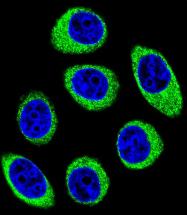 Confocal immunofluorescent analysis of U-251MG cells using ab170812 at a 1/10 dilution, followed by a goat anti-rabbit lgG conjugated to dye (green). DAPI was used to stain the cell nuclei (blue).
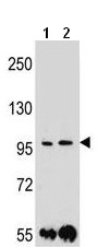 All lanes : Anti-PCDH1 antibody - N-terminal (ab170812) at 1/100 dilutionLane 1 : MDA-MB453 cell line lysatesLane 2 : ZR-75-1 cell line lysatesLysates/proteins at 35 µg per lane.
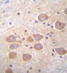 Immunohistochemical analysis of paraffin embedded Human brain tissue labeling PCDH1 with ab170812 at a 1/10 dilution, followed by peroxidase conjugation of the secondary antibody and DAB staining.