![All lanes : Anti-PAX3 antibody [C2] (ab69856) at 1 µg/mlLane 1 : MarkerLane 2 : 624 Mel nuclear lysatedeveloped using the ECL technique](http://www.bioprodhub.com/system/product_images/ab_products/2/sub_4/7683_pax3.gif)
All lanes : Anti-PAX3 antibody [C2] (ab69856) at 1 µg/mlLane 1 : MarkerLane 2 : 624 Mel nuclear lysatedeveloped using the ECL technique
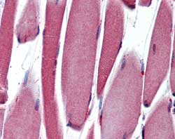
ab109691, at 10 µg/ml, staining PAX3 in formalin-fixed, paraffin-embedded Human Skeletal muscle tissue by Immunohistochemistry.
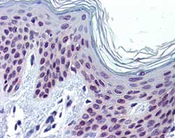
ab109691, at 10 µg/ml, staining PAX3 in formalin-fixed, paraffin-embedded Human Skin tissue by Immunohistochemistry.
![Overlay histogram showing K562 cells stained with ab69856 (red line). The cells were fixed with 80% methanol (5 min) and then permeabilized with 0.1% PBS-Tween for 20 min. The cells were then incubated in 1x PBS / 10% normal goat serum / 0.3M glycine to block non-specific protein-protein interactions followed by the antibody (ab69856, 0.5µg/1x106 cells) for 30 min at 22ºC. The secondary antibody used was DyLight® 488 goat anti-mouse IgG (H+L) (ab96879) at 1/500 dilution for 30 min at 22ºC. Isotype control antibody (black line) was mouse IgG2a [ICIGG2A] (ab91361, 2µg/1x106 cells) used under the same conditions. Acquisition of >5,000 events was performed. This antibody gave a positive signal in K562 cells fixed with 4% paraformaldehyde (10 min)/permeabilized with 0.1% PBS-Tween for 20 min used under the same conditions.](http://www.bioprodhub.com/system/product_images/ab_products/2/sub_4/7686_PAX3-Primary-antibodies-ab69856-3.jpg)
Overlay histogram showing K562 cells stained with ab69856 (red line). The cells were fixed with 80% methanol (5 min) and then permeabilized with 0.1% PBS-Tween for 20 min. The cells were then incubated in 1x PBS / 10% normal goat serum / 0.3M glycine to block non-specific protein-protein interactions followed by the antibody (ab69856, 0.5µg/1x106 cells) for 30 min at 22ºC. The secondary antibody used was DyLight® 488 goat anti-mouse IgG (H+L) (ab96879) at 1/500 dilution for 30 min at 22ºC. Isotype control antibody (black line) was mouse IgG2a [ICIGG2A] (ab91361, 2µg/1x106 cells) used under the same conditions. Acquisition of >5,000 events was performed. This antibody gave a positive signal in K562 cells fixed with 4% paraformaldehyde (10 min)/permeabilized with 0.1% PBS-Tween for 20 min used under the same conditions.