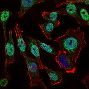
Immunofluorescent analysis of HeLa cells labeling p95 NBS1 with ab181729 at 1/200 (green). Blue: DRAQ5 fluorescent DNA dye. Red: Actin filaments have been labeled with Alexa Fluor-555 phalloidin.
![All lanes : Anti-p95 NBS1 antibody [7E4A2] (ab181729) at 1/500 dilutionLane 1 : A549 cell lysateLane 2 : Jurkat cell lysateLane 3 : PC-12 cell lysate](http://www.bioprodhub.com/system/product_images/ab_products/2/sub_4/6279_ab181729-210936-ab181729.jpg)
All lanes : Anti-p95 NBS1 antibody [7E4A2] (ab181729) at 1/500 dilutionLane 1 : A549 cell lysateLane 2 : Jurkat cell lysateLane 3 : PC-12 cell lysate
![All lanes : Anti-p95 NBS1 antibody [7E4A2] (ab181729) at 1/500 dilutionLane 1 : non-transfected HEK293 cell lysateLane 2 : p95 NBS1 (aa 467-615)-hIgGFc transfected HEK293 cell lysate](http://www.bioprodhub.com/system/product_images/ab_products/2/sub_4/6280_ab181729-210938-ab181729c.jpg)
All lanes : Anti-p95 NBS1 antibody [7E4A2] (ab181729) at 1/500 dilutionLane 1 : non-transfected HEK293 cell lysateLane 2 : p95 NBS1 (aa 467-615)-hIgGFc transfected HEK293 cell lysate
![Anti-p95 NBS1 antibody [7E4A2] (ab181729) at 1/500 dilution + p95 NBS1 (aa 467-615) recombinant protein](http://www.bioprodhub.com/system/product_images/ab_products/2/sub_4/6281_ab181729-210937-ab181729b.jpg)
Anti-p95 NBS1 antibody [7E4A2] (ab181729) at 1/500 dilution + p95 NBS1 (aa 467-615) recombinant protein
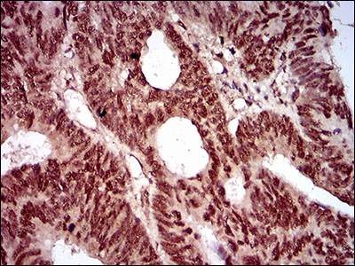
Immunohistochemical analysis of paraffin embedded Human rectum cancer tissue labeling p95 NBS1 with ab181729 at 1/200 with DAB staining.
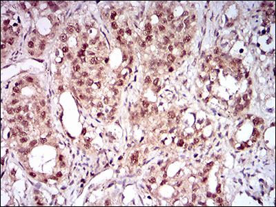
Immunohistochemical analysis of paraffin embedded Human cervical cancer tissue labeling p95 NBS1 with ab181729 at 1/200 with DAB staining.
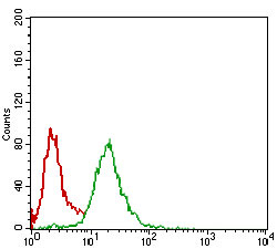
Flow Cytometrical analysis of HeLa cells labeling p95 NBS1 with ab181729 at 1/200 (green) compared to a negative control antibody (red).