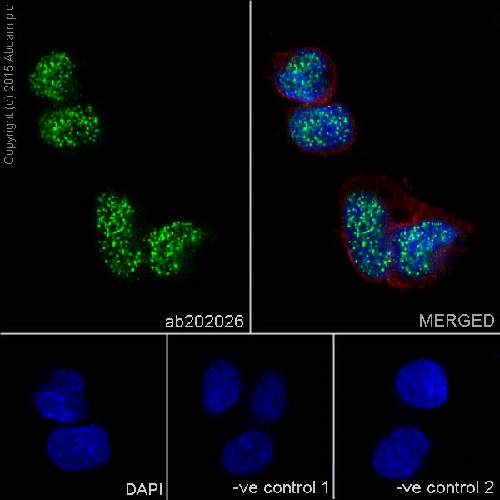
Immunofluorescent analysis of 4% paraformaldehyde-fixed, 0.1% Triton X-100 permeabilized HepG2 (Human liver hepatocellular carcinoma) cells labeling p53 DINP1 with ab202026 at 1/300 dilution, followed by Goat anti-rabbit IgG (Alexa Fluor® 488) (ab150077) secondary antibody at 1/500 dilution (green).Confocal image showing nuclear staining on HepG2 cells.The nuclear counter stain is DAPI (blue).Tubulin is detected with ab7291 (anti-Tubulin mouse mAb) at 1/1000 dilution and ab150120 (AlexaFluor®594 Goat anti-Mouse secondary) at 1/500 dilution (red).The negative controls are as follows:-ve control 1: ab202026 at 1/300 dilution followed by ab150120 (AlexaFluor®594 Goat anti-Mouse secondary) at 1/500 dilution.-ve control 2: ab7291 (anti-Tubulin mouse mAb) at 1/1000 dilution followed by ab150077 (Alexa Fluor®488 Goat Anti-Rabbit IgG H&L) at 1/500 dilution.
![All lanes : Anti-p53 DINP1 antibody [EPR17974] (ab202026) at 1/10000 dilutionLane 1 : HepG2 (Human liver hepatocellular carcinoma) whole cell lysateLane 2 : SK-BR-3 (Human mammary gland adenocarcinoma cell line) whole cell lysateLysates/proteins at 20 µg per lane.SecondaryGoat Anti-Rabbit IgG, (H+L),Peroxidase conjugated at 1/1000 dilution](http://www.bioprodhub.com/system/product_images/ab_products/2/sub_4/5960_ab202026-243844-202026WBa.jpg)
All lanes : Anti-p53 DINP1 antibody [EPR17974] (ab202026) at 1/10000 dilutionLane 1 : HepG2 (Human liver hepatocellular carcinoma) whole cell lysateLane 2 : SK-BR-3 (Human mammary gland adenocarcinoma cell line) whole cell lysateLysates/proteins at 20 µg per lane.SecondaryGoat Anti-Rabbit IgG, (H+L),Peroxidase conjugated at 1/1000 dilution
![Anti-p53 DINP1 antibody [EPR17974] (ab202026) at 1/10000 dilution + Human pancreas lysate at 10 µgSecondaryGoat Anti-Rabbit IgG, (H+L),Peroxidase conjugated at 1/1000 dilution](http://www.bioprodhub.com/system/product_images/ab_products/2/sub_4/5961_ab202026-243843-202026WBb.jpg)
Anti-p53 DINP1 antibody [EPR17974] (ab202026) at 1/10000 dilution + Human pancreas lysate at 10 µgSecondaryGoat Anti-Rabbit IgG, (H+L),Peroxidase conjugated at 1/1000 dilution
![All lanes : Anti-p53 DINP1 antibody [EPR17974] (ab202026) at 1/2000 dilutionLane 1 : C6 (Rat glial tumor cells) whole cell lysateLane 2 : RAW 264.7 (Mouse macrophage cells transformed with Abelson murine leukemia virus) whole cell lysateLane 3 : PC-12 (Rat adrenal gland pheochromocytoma) whole cell lysateLane 4 : NIH/3T3 (Mouse embyro fibroblast cells) whole cell lysateLysates/proteins at 10 µg per lane.SecondaryGoat Anti-Rabbit IgG, (H+L),Peroxidase conjugated at 1/1000 dilution](http://www.bioprodhub.com/system/product_images/ab_products/2/sub_4/5962_ab202026-243842-202026WBc.jpg)
All lanes : Anti-p53 DINP1 antibody [EPR17974] (ab202026) at 1/2000 dilutionLane 1 : C6 (Rat glial tumor cells) whole cell lysateLane 2 : RAW 264.7 (Mouse macrophage cells transformed with Abelson murine leukemia virus) whole cell lysateLane 3 : PC-12 (Rat adrenal gland pheochromocytoma) whole cell lysateLane 4 : NIH/3T3 (Mouse embyro fibroblast cells) whole cell lysateLysates/proteins at 10 µg per lane.SecondaryGoat Anti-Rabbit IgG, (H+L),Peroxidase conjugated at 1/1000 dilution
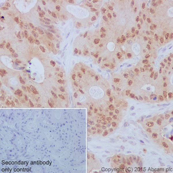
Immunohistochemical analysis of paraffin-embedded Human colonic adenocarcinoma tissue labeling p53 DINP1 with ab202026 at 1/150 dilution, followed by Goat Anti-Rabbit IgG H&L (HRP) (ab97051) secondary antibody at 1/500 dilution.Cytoplasm and nucleus staining on cancer cells of Human colonic adenocarcinoma is observed.Counter stained with Hematoxylin.Secondary antibody only control: Used PBS instead of primary antibody, secondary antibody is Goat Anti-Rabbit IgG H&L (HRP) (ab97051) at 1/500 dilution.
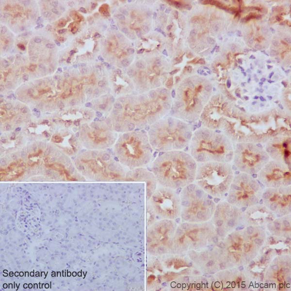
Immunohistochemical analysis of paraffin-embedded Mouse kidney tissue labeling p53 DINP1 with ab202026 at 1/150 dilution, followed by Goat Anti-Rabbit IgG H&L (HRP) (ab97051) secondary antibody at 1/500 dilution.Cytoplasm staining on epithelial cells of mouse kidney tubules is observed.Counter stained with Hematoxylin.Secondary antibody only control: Used PBS instead of primary antibody, secondary antibody is Goat Anti-Rabbit IgG H&L (HRP) (ab97051) at 1/500 dilution.
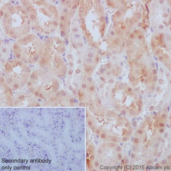
Immunohistochemical analysis of paraffin-embedded Rat kidney tissue labeling p53 DINP1 with ab202026 at 1/150 dilution, followed by Goat Anti-Rabbit IgG H&L (HRP) (ab97051) secondary antibody at 1/500 dilution.Cytoplasm and weak nucleus staining on epithelial cells of rat kidney tubules is observed.Counter stained with Hematoxylin.Secondary antibody only control: Used PBS instead of primary antibody, secondary antibody is Goat Anti-Rabbit IgG H&L (HRP) (ab97051) at 1/500 dilution.