Anti-Nova1 antibody
| Name | Anti-Nova1 antibody |
|---|---|
| Supplier | Abcam |
| Catalog | ab115732 |
| Prices | $407.00 |
| Sizes | 50 µg |
| Host | Goat |
| Clonality | Polyclonal |
| Isotype | IgG |
| Applications | WB ELISA IHC-P |
| Species Reactivities | Human, Mouse, Rat, Monkey |
| Antigen | Synthetic peptide: C-REMPQNVAKTEPVS , corresponding to internal sequence amino acids 124-137 of Human Nova1 Run BLAST with Run BLAST with |
| Description | Goat Polyclonal |
| Gene | NOVA1 |
| Conjugate | Unconjugated |
| Supplier Page | Shop |
Product images
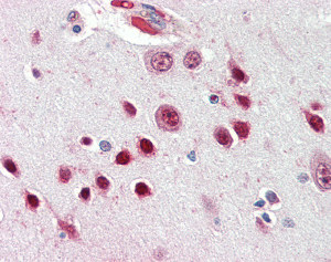 ab115732 at 2.5ug/ml staining Nova1 in Human brain (cortex) tissue by immunohistochemistry (FFPE). Following primary incubation slides were incubated with biotinylated anti-goat IgG secondary antibody, alkaline phosphatase-streptavidin and chromogen.
 ab115732 at 2.5ug/ml staining Nova1 in Human heart tissue by immunohistochemistry (FFPE). Following primary incubation slides were incubated with biotinylated anti-goat IgG secondary antibody, alkaline phosphatase-streptavidin and chromogen.
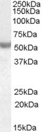 Anti-Nova1 antibody (ab115732) at 0.01 µg/ml + Human breast cancer lysate (in RIPA buffer) at 35 µgdeveloped using the ECL technique