Anti-nNOS (neuronal) (phospho S847) antibody
| Name | Anti-nNOS (neuronal) (phospho S847) antibody |
|---|---|
| Supplier | Abcam |
| Catalog | ab16650 |
| Prices | $392.00 |
| Sizes | 100 µg |
| Host | Rabbit |
| Clonality | Polyclonal |
| Isotype | IgG |
| Applications | IHC-F IHC-F WB |
| Species Reactivities | Mouse, Rat, Fish, Rabbit, Human, Xenopus, Zebrafish |
| Antigen | Synthetic peptide conjugated to KLH derived from within residues 800 - 900 of Mouse nNOS (neuronal), phosphorylated at S847 |
| Blocking Peptide | Mouse nNOS (neuronal) (phospho S847) peptide |
| Description | Rabbit Polyclonal |
| Gene | NOS1 |
| Conjugate | Unconjugated |
| Supplier Page | Shop |
Product images
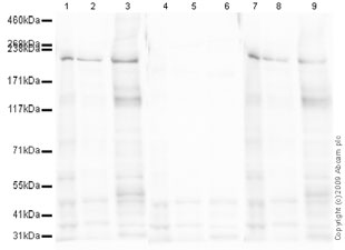 All lanes : Anti-nNOS (neuronal) (phospho S847) antibody (ab16650) at 1 µg/mlLane 1 : Forebrain (Mouse) Tissue LysateLane 2 : Mouse Cortex Tissue LysateLane 3 : Spinal Cord (Mouse) Tissue LysateLane 4 : Forebrain (Mouse) Tissue Lysate with Mouse nNOS (neuronal) (phospho S847) peptide (ab16981) at 1 µg/mlLane 5 : Mouse Cortex Tissue Lysate with Mouse nNOS (neuronal) (phospho S847) peptide (ab16981) at 1 µg/mlLane 6 : Spinal Cord (Mouse) Tissue Lysate with Mouse nNOS (neuronal) (phospho S847) peptide (ab16981) at 1 µg/mlLane 7 : Forebrain (Mouse) Tissue Lysate with nNOS (neuronal) peptide at 1 µg/mlLane 8 : Mouse Cortex Tissue Lysate with nNOS (neuronal) peptide at 1 µg/mlLane 9 : Spinal Cord (Mouse) Tissue Lysate with nNOS (neuronal) peptide at 1 µg/mlLysates/proteins at 20 µg per lane.SecondaryGoat polyclonal to Rabbit IgG - H&L - Pre-Adsorbed (HRP)Performed under reducing conditions.
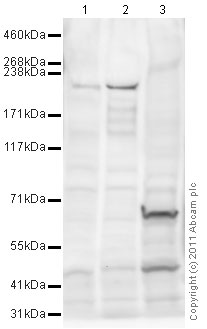 All lanes : Anti-nNOS (neuronal) (phospho S847) antibody (ab16650) at 1 µg/mlLane 1 : Forebrain (Mouse) Tissue LysateLane 2 : Spinal Cord (Mouse) Tissue Lysate Lane 3 : HeLa (Human epithelial carcinoma cell line) Whole Cell Lysate (negative control)Lysates/proteins at 20 µg per lane.SecondaryGoat Anti-Rabbit IgG H&L (HRP) preadsorbed (ab97080) at 1/5000 dilutiondeveloped using the ECL techniquePerformed under reducing conditions.
 Lanes 1 - 3 : Anti-nNOS (neuronal) (phospho S847) antibody (ab16650) at 1 µg/mlLane 4 : nNOS antibody at 1/2500 dilutionLane 1 : mouse forebrainLane 2 : mouse forebrain with Mouse nNOS (neuronal) (phospho S847) peptide (ab16981) at 1 µg/mlLane 3 : mouse forebrain with corresponding unmodified nNOS (neuronal) peptide at 1 µg/mlLane 4 : mouse forebrainLysates/proteins at 20 µg per lane.
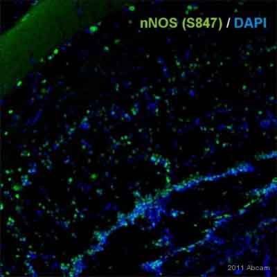
 Immunostaining using Rabbit polyclonal to nNOS (neuronal) (phospho S847) (ab16650) on rat brain tissue sections (30 micron free floating). ab16650 was used at a dilution of 1/3000 and incubated for 18 hours at RT (in PBS triton 0.3%). Secondary Antibody Goat anti-rabbit alexa Fluor 488 was used at a dilution of 1/1000. The image shows cytoplasmic staining of CNS neurons with ab16650 in naïve rats; the staining being observed in the soma and processes of these neurons. The staining was quenched by pre-incubation with peptide against phospho S847 (ab16981), but not by the control peptide (ab57047) indicating that ab16650 is specific for nNos phosphorylated at S847. Protocol: Rats were perfused-fixed with 4% paraformaldehyde. Tissues were post-fixed overnight in the same fixative and then cryoprotected in 20% sucrose overnight. Following embedding in OCT and freezing, tissues were cut and immunostained using the 'free floating’ technique.
 ab16650 staining nNOS (neuronal) (phospho S847) in 16 µm thick sections of Apteronotus leptorhynchus by Immunohistochemistry (Frozen sections).Tissue was fixed in 2% paraformaldehyde, permeabilized using 0.3% Triton X-100, blocked with 3% sheep serum, 1% BSA, 1% teleostean gelatine in TBS for 1 hour at 24°C and then incubated with ab16650 at a 1/100 dilution for 18 hours at 4°C. The secondary used was an Alexa-Fluor 488 conjugated goat anti-rabbit polyclonal used at a 1/200 dilution.See Abreview
Product References
beta3-adrenoreceptor stimulation protects against myocardial infarction injury - beta3-adrenoreceptor stimulation protects against myocardial infarction injury
Niu X, Zhao L, Li X, Xue Y, Wang B, Lv Z, Chen J, Sun D, Zheng Q. PLoS One. 2014 Jun 9;9(6):e98713.
RGSZ2 binds to the neural nitric oxide synthase PDZ domain to regulate mu-opioid - RGSZ2 binds to the neural nitric oxide synthase PDZ domain to regulate mu-opioid
Garzon J, Rodriguez-Munoz M, Vicente-Sanchez A, Bailon C, Martinez-Murillo R, Sanchez-Blazquez P. Antioxid Redox Signal. 2011 Aug 15;15(4):873-87.
Role of neuronal nitric-oxide synthase in estrogen-induced relaxation in rat - Role of neuronal nitric-oxide synthase in estrogen-induced relaxation in rat
Lekontseva O, Chakrabarti S, Jiang Y, Cheung CC, Davidge ST. J Pharmacol Exp Ther. 2011 Nov;339(2):367-75.
Sigma receptor ligand 4-phenyl-1-(4-phenylbutyl)-piperidine modulates neuronal - Sigma receptor ligand 4-phenyl-1-(4-phenylbutyl)-piperidine modulates neuronal
Yang ZJ, Carter EL, Torbey MT, Martin LJ, Koehler RC. Exp Neurol. 2010 Jan;221(1):166-74.
Prolactin promotes oxytocin and vasopressin release by activating neuronal nitric - Prolactin promotes oxytocin and vasopressin release by activating neuronal nitric
Vega C, Moreno-Carranza B, Zamorano M, Quintanar-Stephano A, Mendez I, Thebault S, Martinez de la Escalera G, Clapp C. Am J Physiol Regul Integr Comp Physiol. 2010 Dec;299(6):R1701-8. doi:
Mu-opioid receptors transiently activate the Akt-nNOS pathway to produce - Mu-opioid receptors transiently activate the Akt-nNOS pathway to produce
Sanchez-Blazquez P, Rodriguez-Munoz M, Garzon J. PLoS One. 2010 Jun 23;5(6):e11278.
Coupling between neuronal nitric oxide synthase and glutamate receptor 6-mediated - Coupling between neuronal nitric oxide synthase and glutamate receptor 6-mediated
Yu HM, Xu J, Li C, Zhou C, Zhang F, Han D, Zhang GY. Neuroscience. 2008 Sep 9;155(4):1120-32.