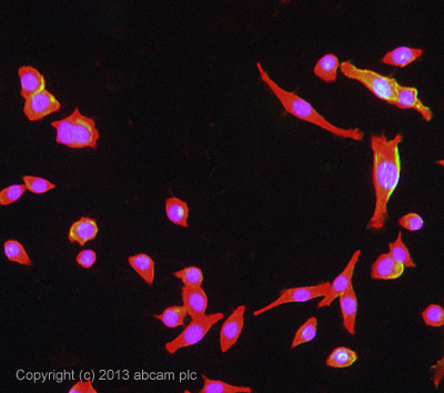
ICC/IF image of ab129051 stained MALME-3M cells. The cells were 100% methanol fixed (5 min) and then incubated in 1%BSA / 10% normal goat serum / 0.3M glycine in 0.1% PBS-Tween for 1h to permeabilise the cells and block non-specific protein-protein interactions. The cells were then incubated with the antibody ab129051 at 5µg/ml overnight at +4°C. The secondary antibody (green) was DyLight® 488 goat anti- rabbit (ab96899) IgG (H+L) used at a 1/250 dilution for 1h. Alexa Fluor® 594 WGA was used to label plasma membranes (red) at a 1/200 dilution for 1h. DAPI was used to stain the cell nuclei (blue) at a concentration of 1.43µM.

IHC-P image of NG2 staining of human melanoma cells in mouse tissue sections using ab129051 (1: 500). The sections were deparaffinized and subjected to heat mediated antigen retrieval using citric acid. The sections were then blocked using 1% BSA for 10 mins at 21°C. ab129051 was dikuted 1:500 using TBS containing BSA and Azide, incubated on the sections for 2 hours at 21°C. The secondary antibody used was Goat polyclonal to Rabbit IgG conjugated to Biotin (1:250).See Abreview

All lanes : Anti-NG2 antibody (ab129051) at 1 µg/ml (Milk blocking - 3%)Lane 1 : Brain (Mouse) Tissue LysateLane 2 : Mouse Hippocampus Tissue LysateLane 3 : Cerebellum Mouse Tissue Lysate Lane 4 : Spinal Cord (Mouse) Tissue LysateLane 5 : Brain (Rat) Tissue LysateLane 6 : Spinal Cord (Rat) Tissue LysateLysates/proteins at 25 µg per lane.SecondaryGoat Anti-Rabbit IgG H&L (HRP) (ab97051) at 1/10000 dilutiondeveloped using the ECL techniquePerformed under reducing conditions.