Anti-NG2 antibody
| Name | Anti-NG2 antibody |
|---|---|
| Supplier | Abcam |
| Catalog | ab83508 |
| Prices | $392.00 |
| Sizes | 100 µg |
| Host | Mouse |
| Clonality | Monoclonal |
| Isotype | IgG1 |
| Applications | IHC-P WB IP ELISA FC ICC/IF ICC/IF IHC-F |
| Species Reactivities | Mouse, Human |
| Antigen | Cell membrane preparation from Human malignant melanoma SK-MEL-28 |
| Description | Mouse Monoclonal |
| Gene | CSPG4 |
| Conjugate | Unconjugated |
| Supplier Page | Shop |
Product images
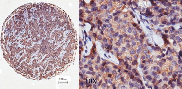 Immunohistochemistry (Formalin/PFA-fixed paraffin-embedded sections) analysis of human breast cancer tissue sections labelling NG2 with ab83508 at 1/50. 4μm-thick sections were deparaffinized in xylene, dehydrated through three alcohol changes. Endogenous peroxidase activity was quenched with 3% hydrogen peroxide in methanol. Antigen retrieval was performed in 96°C solution of 0.01 mol/L sodium citrate buffer (pH 6.0) for 30 minutes. Peroxidase/DAB, Rabbit/Mouse kit, after visualization, the sections were counterstained with hematoxylin. Hisg NG2 expression.
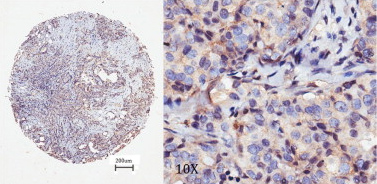 Immunohistochemistry (Formalin/PFA-fixed paraffin-embedded sections) analysis of human breast cancer tissue sections labelling NG2 with ab83508 at 1/50. 4μm-thick sections were deparaffinized in xylene, dehydrated through three alcohol changes. Endogenous peroxidase activity was quenched with 3% hydrogen peroxide in methanol. Antigen retrieval was performed in 96°C solution of 0.01 mol/L sodium citrate buffer (pH 6.0) for 30 minutes. Peroxidase/DAB, Rabbit/Mouse kit, after visualization, the sections were counterstained with hematoxylin. Low NG2 expression.
 Immunohistochemistry (Formalin/PFA-fixed paraffin-embedded sections) analysis of human breast cancer tissue sections labelling NG2 with ab83508 at 1/50. 4μm-thick sections were deparaffinized in xylene, dehydrated through three alcohol changes. Endogenous peroxidase activity was quenched with 3% hydrogen peroxide in methanol. Antigen retrieval was performed in 96°C solution of 0.01 mol/L sodium citrate buffer (pH 6.0) for 30 minutes. Peroxidase/DAB, Rabbit/Mouse kit, after visualization, the sections were counterstained with hematoxylin. Negative NG2 expression.
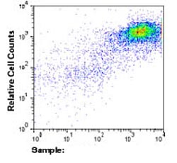 5 x 10⁶ SK-MEL-28 cells were incubated with 5µg of ab83508, followed by APC-GtxMs IgG incubation.
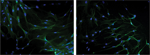 Immunofluorescence analysis of Human adult blood endothelial progenitor cells (left) and umbilical artery smooth muscle cells (right). NG2 was stained using ab83508 at 1/200 dilution. Nuclei were stained with DAPI (blue).
Product References
Endothelial cells from visceral adipose tissue disrupt adipocyte functions in a - Endothelial cells from visceral adipose tissue disrupt adipocyte functions in a
Pellegrinelli V, Rouault C, Veyrie N, Clement K, Lacasa D. Diabetes. 2014 Feb;63(2):535-49.
High chondroitin sulfate proteoglycan 4 expression correlates with poor outcome - High chondroitin sulfate proteoglycan 4 expression correlates with poor outcome
Hsu NC, Nien PY, Yokoyama KK, Chu PY, Hou MF. Biochem Biophys Res Commun. 2013 Nov 15;441(2):514-8. doi:
Unique responses of stem cell-derived vascular endothelial and mesenchymal cells - Unique responses of stem cell-derived vascular endothelial and mesenchymal cells
Keats E, Khan ZA. PLoS One. 2012;7(6):e38752.
Clinicopathological significance of platelet-derived growth factor (PDGF)-B and - Clinicopathological significance of platelet-derived growth factor (PDGF)-B and
Suzuki S, Dobashi Y, Hatakeyama Y, Tajiri R, Fujimura T, Heldin CH, Ooi A. BMC Cancer. 2010 Nov 30;10:659.