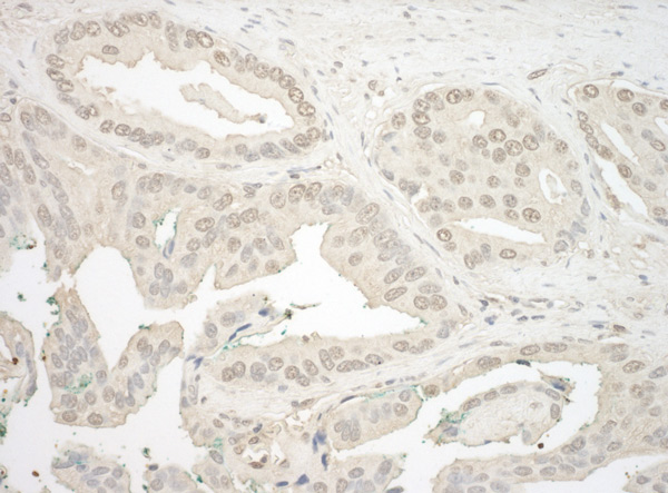
Immunohistochemistry (Formalin/PFA-fixed paraffin-embedded sections) analysis of human prostate carcinoma tissue labelling NFATC4 with ab99431 at 1/1000 (0.2µg/ml). Detection: DAB.
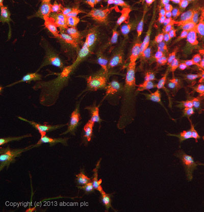
ICC/IF image of ab99431 stained HepG2 cells. The cells were 100% methanol fixed (5 min) and then incubated in 1%BSA / 10% normal goat serum / 0.3M glycine in 0.1% PBS-Tween for 1h to permeabilise the cells and block non-specific protein-protein interactions. The cells were then incubated with the antibody ab99431 at 1µg/ml overnight at +4°C. The secondary antibody (green) was DyLight® 488 goat anti- rabbit (ab96899) IgG (H+L) used at a 1/250 dilution for 1h. Alexa Fluor® 594 WGA was used to label plasma membranes (red) at a 1/200 dilution for 1h. DAPI was used to stain the cell nuclei (blue) at a concentration of 1.43µM.
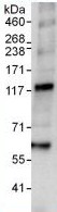
Immunoprecipitation : 1 mg of HeLa cell lysate immunoprecipitated with ab99431 at 3 µg/mg lysate; 20% of immunoprecipitate loaded in lane Western Blot: NFAT3 antibody (ab99431) at 0.4 µg/ml developed using the ECL technique Exposure time : 10 seconds
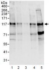
All lanes : Anti-NFATC4 antibody (ab99431) at 0.04 µg/mlLane 1 : HeLa whole cell lysate at 50 µgLane 2 : HeLa whole cell lysate at 15 µgLane 3 : HeLa whole cell lysate at 5 µgLane 4 : 293T whole cell lysate at 50 µgLane 5 : NIH3T3 whole cell lysate at 50 µgdeveloped using the ECL technique