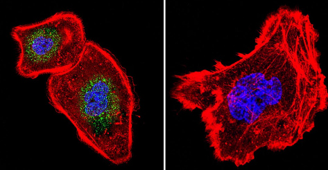
Immunocytochemistry/Immunofluorescence analysis of NFATC4 in A431 cells. Cells were grown on chamber slides and fixed with formaldehyde prior to staining. Cells were probed without (control - right) or with ab3447 at a dilution of 1:100 overnight at 4°C, washed with PBS and incubated with a DyLight-488 conjugated secondary antibody. NFATC4 staining (green), F-Actin staining with Phalloidin (red) and nuclei with DAPI (blue) is shown. Images were taken at 60X magnification.
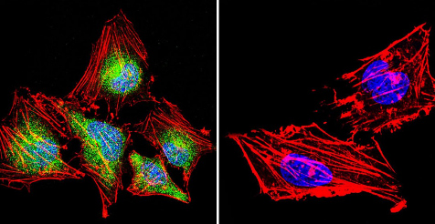
Immunocytochemistry/Immunofluorescence analysis of NFATC4 in HeLa cells. Cells were grown on chamber slides and fixed with formaldehyde prior to staining. Cells were probed without (control - right) or with ab3447 at a dilution of 1:20 overnight at 4°C, washed with PBS and incubated with a DyLight-488 conjugated secondary antibody. NFATC4 staining (green), F-Actin staining with Phalloidin (red) and nuclei with DAPI (blue) is shown. Images were taken at 60X magnification.
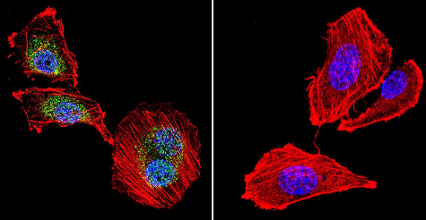
Immunocytochemistry/Immunofluorescence analysis of NFATC4 in U251 cells. Cells were grown on chamber slides and fixed with formaldehyde prior to staining. Cells were incubated without (control - right) or with ab3447 at a dilution of 1:20 overnight at 4°C, washed with PBS and incubated with a DyLight-488 conjugated secondary antibody. NFATC4 staining (green), F-Actin staining with Phalloidin (red) and nuclei with DAPI (blue) is shown. Images were taken at 60X magnification.
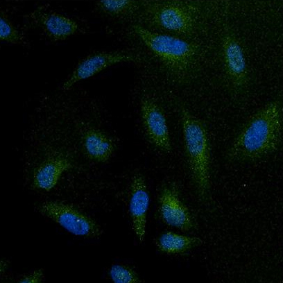
Immunofluorescent analysis of NFATC4 using anti-NFATC4 polyclonal antibody ( ab3447) (shown in green) in HeLa cells. Formalin fixed cells were permeabilized with 0.1% Triton X-100 in TBS for 10 minutes at room temperature. Cells were then blocked with 1% BSA for 15 minutes at room temperature. Cells were probed with a rabbit polyclonal antibody recognizing NFATC4 ( ab3447) at a dilution of 1:100 for at least 1 hour at room temperature. Cells were washed with PBS and incubated with DyLight 488 goat-anti-rabbit secondary antibody at a dilution of 1:400 for 30 minutes at room temperature. Nuclei (blue) were stained with Hoechst 33342 dye. Images were taken at 20X magnification.
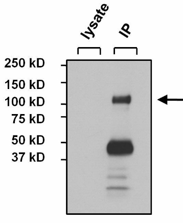
Immunoprecipitation of NFATC4 was performed on MCF7 cells. The antigen:antibody complex was formed by incubating 500µg whole cell lysate with 3µg of rabbit polyclonal antibody recognizing NFATC4 (ab3447) overnight on a rocking platform at 4°C. The immune-complex was captured on 50µl Protein A/G Agarose. Captured immune-complexes were washed and proteins eluted with 5X Reducing Sample Loading Dye . Samples were resolved on a 4-20% Tris-HCl polyacrylamide gel. Proteins were transferred to PVDF membrane and blocked with 5% Milk/TBS-0.1%Tween for at least 1 hour. Membranes were washed in TBS-0.1%Tween 20 and probed with a goat anti-rabbit-HRP secondary antibody at a dilution of 1:20000 for at least one hour. Membranes were washed and chemiluminescent detection performed.
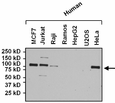
All lanes : Anti-NFATC4 antibody (ab3447) at 1/1000 dilutionLane 1 : MCF7 whole cell lysateLane 2 : Jurkat whole cell lysateLane 3 : Raji whole cell lysateLane 4 : Ramos whole cell lysateLane 5 : HepG2 whole cell lysateLane 6 : U2OS whole cell lysateLane 7 : HeLa whole cell lysateLysates/proteins at 25 µg per lane.SecondaryHRP conjugated Goat anti-rabbit at 1/20000 dilution