Anti-NF-kB p65 antibody
| Name | Anti-NF-kB p65 antibody |
|---|---|
| Supplier | Abcam |
| Catalog | ab16502 |
| Prices | $403.00 |
| Sizes | 100 µg |
| Host | Rabbit |
| Clonality | Polyclonal |
| Isotype | IgG |
| Applications | IHC-F ICC/IF ICC/IF IHC-P WB IP FC |
| Species Reactivities | Mouse, Rat, Chicken, Human, Deer |
| Antigen | Synthetic peptide corresponding to Human NF-kB p65 aa 500 to the C-terminus (C terminal) conjugated to Keyhole Limpet Haemocyanin (KLH) |
| Blocking Peptide | Human NF-kB p65 peptide |
| Description | Rabbit Polyclonal |
| Gene | RELA |
| Conjugate | Unconjugated |
| Supplier Page | Shop |
Product images
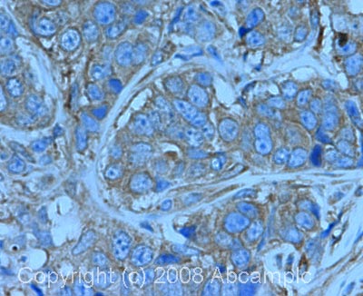 IHC image of NF-kB p65 staining in human breast carcinoma FFPE section, performed on a BondTM system using the standard protocol F. The section was pre-treated using heat mediated antigen retrieval with sodium citrate buffer (pH6, epitope retrieval solution 1) for 20 mins. The section was then incubated with ab16502, 1µg/ml, for 8 mins at room temperature and detected using an HRP conjugated compact polymer system. DAB was used as the chromogen. The section was then counterstained with haematoxylin and mounted with DPX.
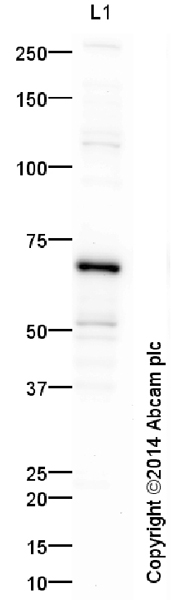 Anti-NF-kB p65 antibody (ab16502) at 1 µg/ml + HeLa (Human epithelial carcinoma cell line) Whole Cell Lysate at 10 µgSecondaryGoat Anti-Rabbit IgG H&L (HRP) (ab97051) at 50000 mg/mldeveloped using the ECL techniquePerformed under reducing conditions.
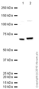 All lanes : Anti-NF-kB p65 antibody (ab16502) at 1 µg/mlLane 1 : Spleen (Mouse) Tissue LysateLane 2 : HeLa (Human epithelial carcinoma cell line) Whole Cell LysateLysates/proteins at 10 µg per lane.SecondaryGoat polyclonal to Rabbit IgG - H&L - Pre-Adsorbed (HRP) at 1/3000 dilutiondeveloped using the ECL techniquePerformed under reducing conditions.
![NF-kB p65 was immunoprecipitated using 0.5mg Hela whole cell extract, 5µg of Rabbit polyclonal to NFkB p65 and 50µl of protein G magnetic beads (+). No antibody was added to the control (-). The antibody was incubated under agitation with Protein G beads for 10min, Hela whole cell extract lysate diluted in RIPA buffer was added to each sample and incubated for a further 10min under agitation.Proteins were eluted by addition of 40µl SDS loading buffer and incubated for 10min at 70oC; 10µl of each sample was separated on a SDS PAGE gel, transferred to a nitrocellulose membrane, blocked with 5% BSA and probed with ab16502.Secondary: Mouse monoclonal [SB62a] Secondary Antibody Anti-Rabbit HRP (IgG light chain) (ab99697).Band: 68kDa: NFkB p65](http://www.bioprodhub.com/system/product_images/ab_products/2/sub_3/29718_NFkB-p65-Primary-antibodies-ab16502-57.jpg) NF-kB p65 was immunoprecipitated using 0.5mg Hela whole cell extract, 5µg of Rabbit polyclonal to NFkB p65 and 50µl of protein G magnetic beads (+). No antibody was added to the control (-). The antibody was incubated under agitation with Protein G beads for 10min, Hela whole cell extract lysate diluted in RIPA buffer was added to each sample and incubated for a further 10min under agitation.Proteins were eluted by addition of 40µl SDS loading buffer and incubated for 10min at 70oC; 10µl of each sample was separated on a SDS PAGE gel, transferred to a nitrocellulose membrane, blocked with 5% BSA and probed with ab16502.Secondary: Mouse monoclonal [SB62a] Secondary Antibody Anti-Rabbit HRP (IgG light chain) (ab99697).Band: 68kDa: NFkB p65
NF-kB p65 was immunoprecipitated using 0.5mg Hela whole cell extract, 5µg of Rabbit polyclonal to NFkB p65 and 50µl of protein G magnetic beads (+). No antibody was added to the control (-). The antibody was incubated under agitation with Protein G beads for 10min, Hela whole cell extract lysate diluted in RIPA buffer was added to each sample and incubated for a further 10min under agitation.Proteins were eluted by addition of 40µl SDS loading buffer and incubated for 10min at 70oC; 10µl of each sample was separated on a SDS PAGE gel, transferred to a nitrocellulose membrane, blocked with 5% BSA and probed with ab16502.Secondary: Mouse monoclonal [SB62a] Secondary Antibody Anti-Rabbit HRP (IgG light chain) (ab99697).Band: 68kDa: NFkB p65
 ab16502 stining the nuclei of the cardiac cells in rat tissue. The tissues were fixed (animals perfused fixed) with 4% PFA and later postfixed overnight in the same fixative. They were cryoprotected in 30% sucrose and cut using a cryostat.See Abreview
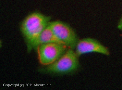 ICC/IF image of ab16502 stained MCF7 cells. The cells were 4% PFA fixed (10 min) and then incubated in 1%BSA / 10% normal goat serum / 0.3M glycine in 0.1% PBS-Tween for 1h to permeabilise the cells and block non-specific protein-protein interactions. The cells were then incubated with the antibody (ab16502, 1µg/ml) overnight at +4°C. The secondary antibody (green) was goat anti-rabbit DyLight® 488 (IgG - H&L, pre-adsorbed) (ab96899) used at a 1/250 dilution for 1h. Alexa Fluor® 594 WGA was used to label plasma membranes (red) at a 1:200 dilution for 1h. DAPI was used to stain the cell nuclei (blue) at a concentration of 1.43µM.
 Performed under reducing conditions.
 Performed under reducing conditions.
 Performed under reducing conditions.
 Performed under reducing conditions.
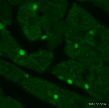
 ab16502 at a 1/500 dilution staining Asynchronous and paraformaldehyde-fixed (4%) HeLa cells by immunocytochemistry. The antibody was incubated with the cells 30 minutes and then detected using a Cy3 conjugated Goat Anti-Mouse IgG (H+L) antibody.This image is courtesy of an Abreview by Kirk McManus submitted on 27 February 2006.See Abreview
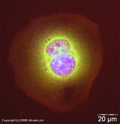 ICC/IF image of ab16502 stained human HeLa cells. The cells were methanol fixed (5 min) and incubated with the antibody (ab16502, 1µg/ml) for 1h at room temperature. The secondary antibody (green) was Alexa Fluor® 488 goat anti-rabbit IgG (H+L) used at a 1/1000 dilution for 1h. Image-iTTM FX Signal Enhancer was used as the primary blocking agent, 5% BSA (in TBS-T) was used for all other blocking steps. DAPI was used to stain the cell nuclei (blue). Alexa Fluor® 594 phalloidin was used to label F-actin (red).
Product References
Clearance of senescent hepatocytes in a neoplastic-prone microenvironment delays - Clearance of senescent hepatocytes in a neoplastic-prone microenvironment delays
Marongiu F, Serra MP, Sini M, Angius F, Laconi E. Aging (Albany NY). 2014 Jan;6(1):26-34.
Overexpression of heat shock protein 72 attenuates NF-kappaB activation using a - Overexpression of heat shock protein 72 attenuates NF-kappaB activation using a
Sheppard PW, Sun X, Khammash M, Giffard RG. PLoS Comput Biol. 2014 Feb 6;10(2):e1003471.
CD31 is a key coinhibitory receptor in the development of immunogenic dendritic - CD31 is a key coinhibitory receptor in the development of immunogenic dendritic
Clement M, Fornasa G, Guedj K, Ben Mkaddem S, Gaston AT, Khallou-Laschet J, Morvan M, Nicoletti A, Caligiuri G. Proc Natl Acad Sci U S A. 2014 Mar 25;111(12):E1101-10. doi:
Nrf2 upregulates ATP binding cassette transporter expression and activity at the - Nrf2 upregulates ATP binding cassette transporter expression and activity at the
Wang X, Campos CR, Peart JC, Smith LK, Boni JL, Cannon RE, Miller DS. J Neurosci. 2014 Jun 18;34(25):8585-93.
A gain-of-function mouse model identifies PRMT6 as a NF-kappaB coactivator. - A gain-of-function mouse model identifies PRMT6 as a NF-kappaB coactivator.
Di Lorenzo A, Yang Y, Macaluso M, Bedford MT. Nucleic Acids Res. 2014 Jul;42(13):8297-309.
Comparative analysis of the cytotoxic effects of okadaic acid-group toxins on - Comparative analysis of the cytotoxic effects of okadaic acid-group toxins on
Ferron PJ, Hogeveen K, Fessard V, Le Hegarat L. Mar Drugs. 2014 Aug 21;12(8):4616-34.
MCPIP1 contributes to the toxicity of proteasome inhibitor MG-132 in HeLa cells - MCPIP1 contributes to the toxicity of proteasome inhibitor MG-132 in HeLa cells
Skalniak L, Dziendziel M, Jura J. Mol Cell Biochem. 2014 Oct;395(1-2):253-63.
Low-dose arsenic induces chemotherapy protection via p53/NF-kappaB-mediated - Low-dose arsenic induces chemotherapy protection via p53/NF-kappaB-mediated
Ganapathy S, Xiao S, Seo SJ, Lall R, Yang M, Xu T, Su H, Shadfan M, Ha CS, Yuan ZM. Oncogene. 2014 Mar 13;33(11):1359-66.
Induction of cytopathogenicity in human glioblastoma cells by chikungunya virus. - Induction of cytopathogenicity in human glioblastoma cells by chikungunya virus.
Abraham R, Mudaliar P, Padmanabhan A, Sreekumar E. PLoS One. 2013 Sep 25;8(9):e75854.
Ezrin-radixin-moesin-binding phosphoprotein 50 (EBP50) and nuclear factor-kappaB - Ezrin-radixin-moesin-binding phosphoprotein 50 (EBP50) and nuclear factor-kappaB
Leslie KL, Song GJ, Barrick S, Wehbi VL, Vilardaga JP, Bauer PM, Bisello A. J Biol Chem. 2013 Dec 20;288(51):36426-36.