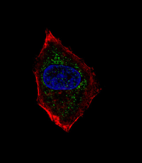
Immunofluorescence of HepG2 cell labelling NeuroD1 with ab76685. HepG2 cells were fixed with 4% PFA (20 min), permeabilized with Triton X-100 (0.1%, 10 min), then incubated with ab76685 (1:25, 1 h at 37℃). Alexa Fluor® 488 conjugated donkey anti-rabbit antibody (green) was used as the secondary antibody (1:400, 50 min at 37℃). Cytoplasmic actin was counterstained with Alexa Fluor® 555 (red) conjugated Phalloidin (7units/ml, 1 h at 37℃) and nuclei were counterstained with DAPI (blue) (10 µg/ml, 10 min). NeuroD1 immunoreactivity is localised to vesicles.

ab76685, at a 1/50 dilution, staining NeuroD1 in formalin fixed, paraffin embedded human hepatocarcinoma tissue by Immunohistochemistry. A peroxidase conjugated secondary antibody was then used, followed by DAB staining.
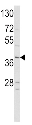 Anti-NeuroD1 antibody (ab76685) at 1/100 dilution + HepG2 cell line lysate at 35 µg