
ab113202 staining NET1 in PC-12 cells treated with milnacipran hydrochloride (ab120755), by ICC/IF. Decrease of NET1 expression correlates with increased concentration of milnacipran hydrochloride, as described in literature.The NGF treated cells were incubated at 37°C for 6 hour in media containing different concentrations of ab120755 (milnacipran hydrochloride ) in DMSO, fixed with 4% formaldehyde for 10 minutes at room temperature and blocked with PBS containing 10% goat serum, 0.3 M glycine, 1% BSA and 0.1% tween for 2h at room temperature. Staining of the treated cells with ab113202 (5 μg/ml) was performed overnight at 4°C in PBS containing 1% BSA and 0.1% tween. A DyLight 488 anti-rabbit polyclonal antibody (ab96899) at 1/250 dilution was used as the secondary antibody. Nuclei were counterstained with DAPI and are shown in blue.
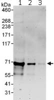
Immunoprecipitation of NET1 (6 µg of antibody per mg of Jurkat cell lysate; 20% of the IP loaded). Lane 1- Immunoprecipitation with an anti-Net1 antibody that recognizes an upstream epitope; Lane 2- Immunoprecipitation with ab113202; Lane 3- Immunoprecipitation with IgG control. Anti-Net 1 antibody (ab113202) used at 1 µg/ml for western blot. Detection: Chemiluminescence with exposure time of 3 min.
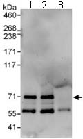
 All lanes : Anti-NET1 antibody (ab113202) at 0.4 µg/mlLane 1 : Jurkat Cell lysate at 50 µgLane 2 : Jurkat Cell lysate at 15 µgLane 3 : Jurkat Cell lysate at 5 µg