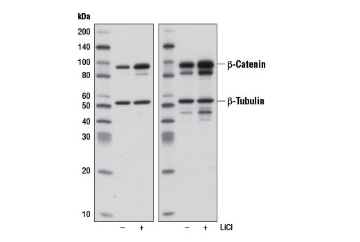
Western blot analysis of extracts from 293T cells, untreated (-) or treated (+) with LiCl (20 mM, 20 hr at 37ºC), using Non-phospho (Active) β-Catenin (Ser33/37/Thr41) (D13A1) Rabbit mAb (left) and β-Catenin Antibody #9562 (right). Equal protein loading was confirmed using β-Tubulin (9F3) Rabbit mAb #2128.
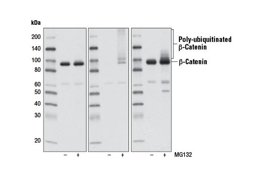
Western blot analysis of extracts from 293T cells, untreated (-) or treated (+) with MG132 (10 μM, 4 hr at 37ºC), using Non-phospho (Active) β-Catenin (Ser33/37/Thr41) (D13A1) Rabbit mAb (left), Phospho-β-Catenin (Ser33/37/Thr41) Antibody #9561 (center), or β-Catenin (6B3) Rabbit mAb #9582 (right). Note that Non-phospho (Active) β-Catenin (Ser33/37/Thr41) (D13A1) Rabbit mAb fails to detect poly-ubiquitinated β-catenin in MG132-treated cells, indicating its specificity for stabilized protein.
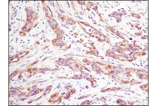
Immunohistochemical analysis of paraffin embedded human breast carcinoma using Non-phospho (Active) β-Catenin (Ser33/37/Thr41) (D13A1) Rabbit mAb.
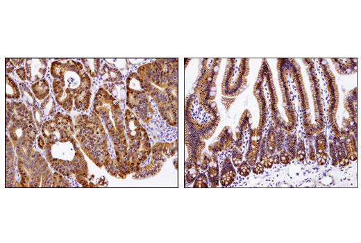
Immunohistochemical analysis of paraffin-embedded Apc (Min/+) mouse intestinal adenoma (left) and adjacent normal intestinal epithelium (right) using Non-phospho (Active) beta-Catenin (Ser33/37/Thr41) (D13A1) Rabbit mAb. Note the nuclear accumulation of active beta-Catenin in the adenoma cells.
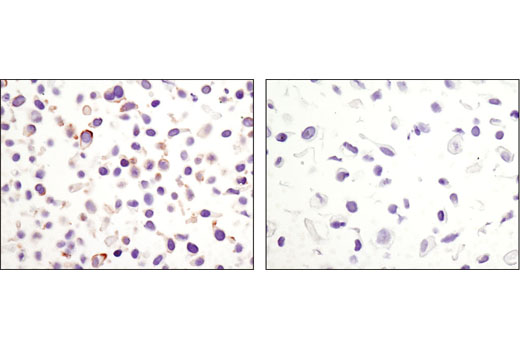
Immunohistochemical analysis of paraffin embedded cell pellets, HeLa (left) or NCI-H28 (right), using Non-phospho (Active) β-Catenin (Ser33/37/Thr41) (D13A1) Rabbit mAb.
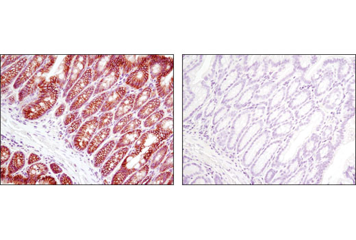
Immunohistochemical analysis of paraffin embedded mouse colon using Non-phospho (Active) β-Catenin (Ser33/37/Thr41) (D13A1) Rabbit mAb in the presence of phospho-β-catenin (Ser33/37/Thr41) peptide (left) or non-phospho-β-catenin (Ser33/37/Thr41) peptide (right). Note the absence of staining only in the presence of the non-phospho-β-catenin (Ser33/37/Thr41) peptide, demonstrating non-phospho specificity.
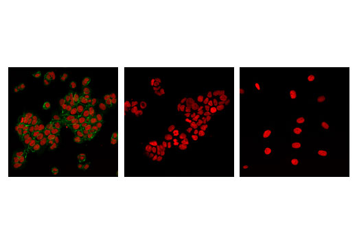
Confocal immunofluorescent analysis of HCT-15 (left, center; positive) or NCI-H28 (right; negative) cells using Non-phospho (Active) β-Catenin (Ser33/37/Thr41) (D13A1) Rabbit mAb (green) in the presence of Phospho-Catenin-β (Ser33/Ser37/Thr41) peptide (left) or Non-phospho-Catenin-β (Ser33/Ser37/Thr41) peptide (center). Red = Propidium Iodide (PI)/RNase Staining Solution #4087.
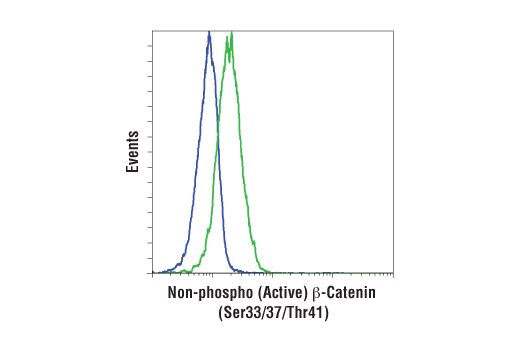
Flow cytometric analysis of K562 cells, untreated (blue) or treated with 6-bromoindirubin-3'-oxime (BIO) (30 nM, 40 hr) (green) using Non-phospho (Active) β-Catenin (Ser33/37/Thr41) (D13A1) Rabbit mAb.
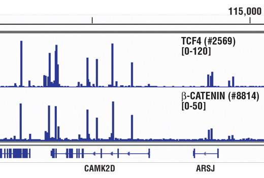
Chromatin immunoprecipitations were performed with cross-linked chromatin from 4 x 10 6 HCT116 cells and either 10 μl of TCF4 (C48H11) Rabbit mAb #2569 or 5 μl of Non-phospho (Active) β-Catenin (Ser33/37/Thr41) (D13A1) Rabbit mAb, using SimpleChIP ® Enzymatic Chromatin IP Kit (Magnetic Beads) #9003. DNA Libraries were prepared from 5ng enriched ChIP DNA using NEBNext ® Ultra™ II DNA Library Prep Kit for Illumina ® , and sequenced on the Illumina NextSeq. TCF4 and β-Catenin are known to associate with each other on chromatin. The figure shows binding of both TCF4 and β-Catenin across CAMK2D, a known target gene of both TCF4 and β-Catenin (see additional figure containing ChIP-qPCR data). For additional ChIP-seq tracks, please download the product data sheet.
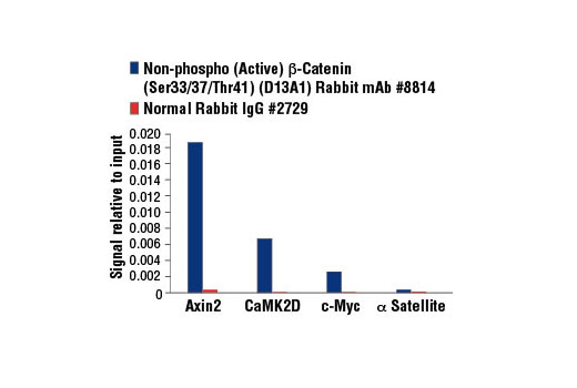
Chromatin immunoprecipitations were performed with cross-linked chromatin from 4 x 10 6 HCT 116 cells and either 5 μl of Non-phospho (Active) β-Catenin (Ser33/37/Thr41) (D13A1) Rabbit mAb or 2 μl of Normal Rabbit IgG #2729 using SimpleChIP ® Enzymatic Chromatin IP Kit (Magnetic Beads) #9003. The enriched DNA was quantified by real-time PCR using SimpleChIP ® Human Axin2 Intron 1 Primers #8973, SimpleChIP ® Human CaMK2D Intron 3 Primers #5111, human c-Myc promoter primers, and SimpleChIP ® Human α Satellite Repeat Primers #4486. The amount of immunoprecipitated DNA in each sample is represented as signal relative to the total amount of input chromatin, which is equivalent to one.


