![Anti-Nestin antibody [SP103] (ab105389) at 1/100 dilution + HeLa cell lysate](http://www.bioprodhub.com/system/product_images/ab_products/2/sub_3/29100_Nestin-Primary-antibodies-ab105389-1.jpg)
Anti-Nestin antibody [SP103] (ab105389) at 1/100 dilution + HeLa cell lysate
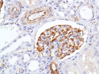
ab105389, at a 1/100 dilution, staining Nestin in formalin fixed, paraffin embedded Human kidney tissue by Immunohistochemistry.
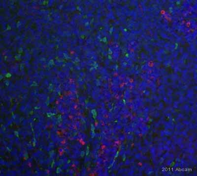
ab105389 staining Nestin in human melanoma tissue by Immunohistochemistry (Formalin/PFA-fixed paraffin-embedded sections).Tissue was fixed in formaldehyde and a heat mediated antigen retrieval step was performed using Tris EDTA pH 9.0. Samples were then permeabilized using 0.25% Triton X-100, blocked, then incubated with ab105389 at a 1/100 dilution for 18 hours at 4°C. The secondary used was an Alexa-Fluor 594 (red) conjugated donkey anti-rabbit polyclonal used at a 1/100 dilution. (Green) tumor cells, (blue) DAPI.See Abreview
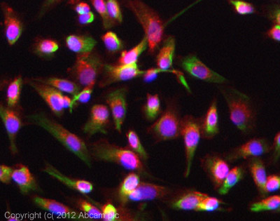
ICC/IF image of ab105389 stained SKNSH cells. The cells were 4% formaldehyde fixed (10 min) and then incubated in 1%BSA / 10% normal goat serum / 0.3M glycine in 0.1% PBS-Tween for 1h to permeabilise the cells and block non-specific protein-protein interactions. The cells were then incubated with the antibody (ab105389, 1/200 dilution) overnight at +4°C. The secondary antibody (green) was ab96899, DyLight® 488 goat anti-rabbit IgG (H+L) used at a 1/250 dilution for 1h. Alexa Fluor® 594 WGA was used to label plasma membranes (red) at a 1/200 dilution for 1h. DAPI was used to stain the cell nuclei (blue) at a concentration of 1.43µM.
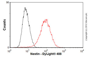
Overlay histogram showing SH-SY5Y cells stained with ab105389 (red line). The cells were fixed with 4% paraformaldehyde (10 min) and then permeabilized with 0.1% PBS-Tween for 20 min. The cells were then incubated in 1x PBS / 10% normal goat serum / 0.3M glycine to block non-specific protein-protein interactions followed by the antibody (ab105389, 1/50 dilution) for 30 min at 22ºC. The secondary antibody used was DyLight® 488 goat anti-rabbit IgG (H+L) (ab96899) at 1/500 dilution for 30 min at 22ºC. Isotype control antibody (black line) was rabbit IgG (monoclonal) (1µg/1x106 cells) used under the same conditions. Acquisition of >5,000 events was performed.