Anti-Nestin antibody [2C1.3A11]
| Name | Anti-Nestin antibody [2C1.3A11] |
|---|---|
| Supplier | Abcam |
| Catalog | ab18102 |
| Prices | $388.00 |
| Sizes | 100 µl |
| Host | Mouse |
| Clonality | Monoclonal |
| Isotype | IgG1 |
| Clone | 2C1.3A11 |
| Applications | WB ICC/IF FC IHC-P IP |
| Species Reactivities | Human |
| Antigen | A 150 amino acid fragment from the cloned human Nestin |
| Description | Mouse Monoclonal |
| Gene | NES |
| Conjugate | Unconjugated |
| Supplier Page | Shop |
Product images
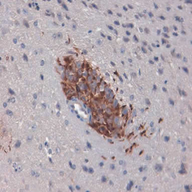 Immunohistochemistry (Formalin/PFA-fixed paraffin-embedded sections) analysis of migrating tumor cells labelling Nestin with ab18102 at 2µg/ml. Cytoplasm and membrane positive staining shown.
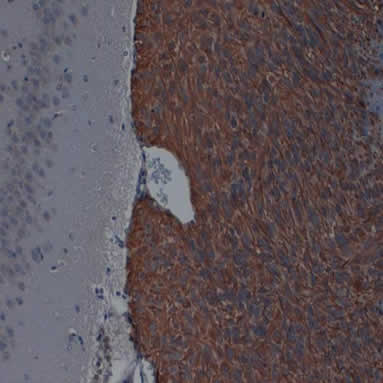 Immunohistochemistry (Formalin/PFA-fixed paraffin-embedded sections) analysis of xenograft tumor cells labelling Nestin with ab18102.
![All lanes : Anti-Nestin antibody [2C1.3A11] (ab18102) at 1/1000 dilutionLane 1 : Total rat brain protein at 5 µgLane 2 : Human CNS progenitor cell protein at 1 µgLane 3 : Human CNS progenitor cell protein at 5 µg](http://www.bioprodhub.com/system/product_images/ab_products/2/sub_3/29076_ab18102-219266-ab18102wb.jpg) All lanes : Anti-Nestin antibody [2C1.3A11] (ab18102) at 1/1000 dilutionLane 1 : Total rat brain protein at 5 µgLane 2 : Human CNS progenitor cell protein at 1 µgLane 3 : Human CNS progenitor cell protein at 5 µg
All lanes : Anti-Nestin antibody [2C1.3A11] (ab18102) at 1/1000 dilutionLane 1 : Total rat brain protein at 5 µgLane 2 : Human CNS progenitor cell protein at 1 µgLane 3 : Human CNS progenitor cell protein at 5 µg
![Overlay histogram showing SH-SY5Y cells stained with ab18102 (red line). The cells were fixed with 4% paraformaldehyde (10 min) and then permeabilized with 0.1% PBS-Tween for 20 min. The cells were then incubated in 1x PBS / 10% normal goat serum / 0.3M glycine to block non-specific protein-protein interactions followed by the antibody (ab18102, 1/200 dilution) for 30 min at 22ºC. The secondary antibody used was DyLight® 488 goat anti-mouse IgG (H+L) (ab96879) at 1/500 dilution for 30 min at 22ºC. Isotype control antibody (black line) was mouse IgG1 [ICIGG1] (ab91353, 2µg/1x106 cells) used under the same conditions. Acquisition of >5,000 events was performed.](http://www.bioprodhub.com/system/product_images/ab_products/2/sub_3/29077_Nestin-Primary-antibodies-ab18102-1.jpg) Overlay histogram showing SH-SY5Y cells stained with ab18102 (red line). The cells were fixed with 4% paraformaldehyde (10 min) and then permeabilized with 0.1% PBS-Tween for 20 min. The cells were then incubated in 1x PBS / 10% normal goat serum / 0.3M glycine to block non-specific protein-protein interactions followed by the antibody (ab18102, 1/200 dilution) for 30 min at 22ºC. The secondary antibody used was DyLight® 488 goat anti-mouse IgG (H+L) (ab96879) at 1/500 dilution for 30 min at 22ºC. Isotype control antibody (black line) was mouse IgG1 [ICIGG1] (ab91353, 2µg/1x106 cells) used under the same conditions. Acquisition of >5,000 events was performed.
Overlay histogram showing SH-SY5Y cells stained with ab18102 (red line). The cells were fixed with 4% paraformaldehyde (10 min) and then permeabilized with 0.1% PBS-Tween for 20 min. The cells were then incubated in 1x PBS / 10% normal goat serum / 0.3M glycine to block non-specific protein-protein interactions followed by the antibody (ab18102, 1/200 dilution) for 30 min at 22ºC. The secondary antibody used was DyLight® 488 goat anti-mouse IgG (H+L) (ab96879) at 1/500 dilution for 30 min at 22ºC. Isotype control antibody (black line) was mouse IgG1 [ICIGG1] (ab91353, 2µg/1x106 cells) used under the same conditions. Acquisition of >5,000 events was performed.
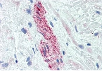 Immunohistochemistry of Anti-Nestin on formalin fixed paraffin embedded prostate nerve tissue using ab18102
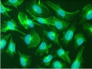 Immunofluorescence staining of U251 cells (glioblastoma cell line) stained using ab18102(green) and bisbenzimide (blue)
Product References
STAT3 is required for proliferation and maintenance of multipotency in - STAT3 is required for proliferation and maintenance of multipotency in
Sherry MM, Reeves A, Wu JK, Cochran BH. Stem Cells. 2009 Oct;27(10):2383-92.
Coexpression of nestin in neural and glial cells in the developing human CNS - Coexpression of nestin in neural and glial cells in the developing human CNS
Messam CA, Hou J, Major EO. Exp Neurol. 2000 Feb;161(2):585-96.