![Overlay histogram showing HepG2 cells stained with ab110249 (red line). The cells were fixed with 80% methanol (5 min) and then permeabilized with 0.1% PBS-Tween for 20 min. The cells were then incubated in 1x PBS / 10% normal goat serum / 0.3M glycine to block non-specific protein-protein interactions followed by the antibody (ab110249, 1μg/1x106 cells) for 30 min at 22°C. The secondary antibody used was Alexa Fluor® 488 goat anti-mouse IgG (H&L) (ab150113) at 1/2000 dilution for 30 min at 22°C. Isotype control antibody (black line) was mouse IgG1 [ICIGG1] (ab91353, 1μg/1x106 cells) used under the same conditions. Unlabelled sample (blue line) was also used as a control. Acquisition of >5,000 events were collected using a 20mW Argon ion laser (488nm) and 525/30 bandpass filter.](http://www.bioprodhub.com/system/product_images/ab_products/2/sub_3/28598_ab110249-3-ab110249FC.jpg)
Overlay histogram showing HepG2 cells stained with ab110249 (red line). The cells were fixed with 80% methanol (5 min) and then permeabilized with 0.1% PBS-Tween for 20 min. The cells were then incubated in 1x PBS / 10% normal goat serum / 0.3M glycine to block non-specific protein-protein interactions followed by the antibody (ab110249, 1μg/1x106 cells) for 30 min at 22°C. The secondary antibody used was Alexa Fluor® 488 goat anti-mouse IgG (H&L) (ab150113) at 1/2000 dilution for 30 min at 22°C. Isotype control antibody (black line) was mouse IgG1 [ICIGG1] (ab91353, 1μg/1x106 cells) used under the same conditions. Unlabelled sample (blue line) was also used as a control. Acquisition of >5,000 events were collected using a 20mW Argon ion laser (488nm) and 525/30 bandpass filter.
![All lanes : Anti-NDUFS2 antibody [7A12BE5AD5] (ab110249) at 0.25 µg/mlLane 1 : Molecular Weight Markers Lane 2 : Isolated mitochondria from Human liverLane 3 : Isolated mitochondria from Bovine heart Lane 4 : Isolated mitochondria from H9C2 cells (Rat cardiomyocyte) Lane 5 : Isolated mitochondria from MEF cells (Mouse embryo fibroblast) Lane 6 : Isolated mitochondria from HepG2](http://www.bioprodhub.com/system/product_images/ab_products/2/sub_3/28599_testtesttest-Primary-antibodies-ab110249-1.jpg)
All lanes : Anti-NDUFS2 antibody [7A12BE5AD5] (ab110249) at 0.25 µg/mlLane 1 : Molecular Weight Markers Lane 2 : Isolated mitochondria from Human liverLane 3 : Isolated mitochondria from Bovine heart Lane 4 : Isolated mitochondria from H9C2 cells (Rat cardiomyocyte) Lane 5 : Isolated mitochondria from MEF cells (Mouse embryo fibroblast) Lane 6 : Isolated mitochondria from HepG2
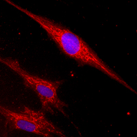
Mitochondrial localization of Complex I subunit NDUFS2 visualized by Immunocytochemistry in HDFn cultured cells (normal Human dermal fibroblasts, neonatal), using ab110249 at 5 µg/ml.