Anti-Ndufs1 antibody [EPR11521(B)] (Alexa Fluor® 647)
| Name | Anti-Ndufs1 antibody [EPR11521(B)] (Alexa Fluor® 647) |
|---|---|
| Supplier | Abcam |
| Catalog | ab198956 |
| Prices | $425.00 |
| Sizes | 100 µl |
| Host | Rabbit |
| Clonality | Monoclonal |
| Isotype | IgG |
| Clone | EPR11521(B) |
| Applications | ICC/IF ICC/IF |
| Species Reactivities | Mouse, Rat, Human |
| Antigen | Synthetic peptide (the amino acid sequence is considered to be commercially sensitive) corresponding to Human Ndufs1 |
| Description | Rabbit Monoclonal |
| Gene | NDUFS1 |
| Conjugate | Alexa Fluor® 647 |
| Supplier Page | Shop |
Product images
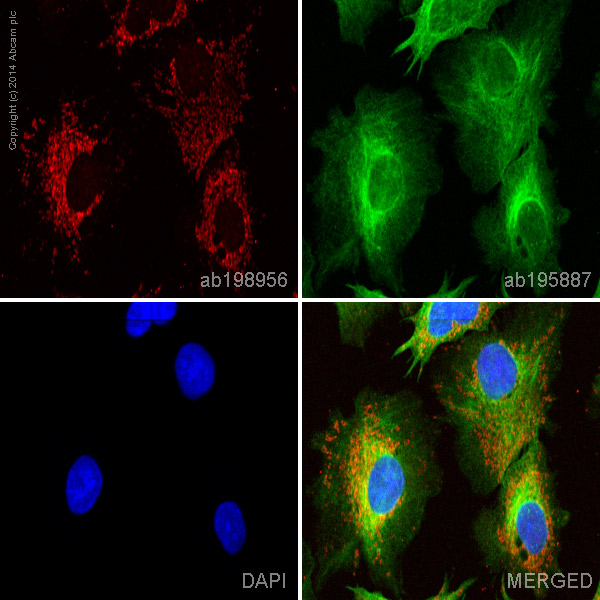 ab198956 staining Ndufs1 in HepG2 cells. The cells were fixed with 100% methanol (5 min), permeabilized with 0.1% Triton X-100 for 5 minutes and then blocked with 1% BSA/10% normal goat serum/0.3M glycine in 0.1% PBS-Tween for 1h. The cells were then incubated overnight at +4°C with ab198956 at 1/100 dilution (shown in red) and ab195887, Mouse monoclonal to alpha Tubulin (Alexa Fluor® 488), at 1/250 dilution (shown in green). Nuclear DNA was labelled with DAPI (shown in blue).Image was taken with a confocal microscope (Leica-Microsystems, TCS SP8).