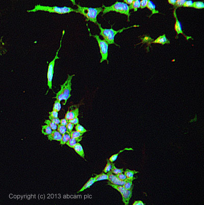
ICC/IF image of ab33164 stained F9 cells. The cells were 4% formaldehyde fixed (10 min) and then incubated in 1%BSA / 10% normal goat serum / 0.3M glycine in 0.1% PBS-Tween for 1h to permeabilise the cells and block non-specific protein-protein interactions. The cells were then incubated with the antibody ab33164 at 5µg/ml overnight at +4°C. The secondary antibody (green) was DyLight® 488 goat anti- rabbit (ab96899) IgG (H+L) used at a 1/250 dilution for 1h. Alexa Fluor® 594 WGA was used to label plasma membranes (red) at a 1/200 dilution for 1h. DAPI was used to stain the cell nuclei (blue) at a concentration of 1.43µM.
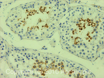
IHC image of MSY2/BOX2 staining in human testis formalin fixed paraffin embedded tissue section, performed on a Leica BondTM system using the standard protocol F. The section was pre-treated using heat mediated antigen retrieval with sodium citrate buffer (pH6, epitope retrieval solution 1) for 20 mins. The section was then incubated with ab33164, 1µg/ml, for 15 mins at room temperature and detected using an HRP conjugated compact polymer system. DAB was used as the chromogen. The section was then counterstained with haematoxylin and mounted with DPX.
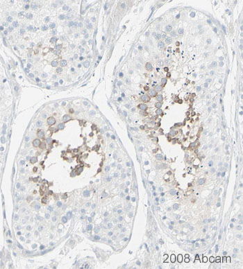
Image courtesy of Human Protein Atlasab33164 staining MSY2/YBOX2 in human testis tissue, showing staining in the seminiferous tubules. Paraffin embedded human skin tissue was incubated with ab33164 (1/200 dilution) for 30 mins at room temperature. Antigen retrieval was performed by heat induction in citrate buffer pH 6. ab33164 was tested in a tissue microarray (TMA) containing a wide range of normal and cancer tissues as well as a cell microarray consisting of a range of commonly used, well characterised human cell lines. Further results for this antibody can be found at www.proteinatlas.org
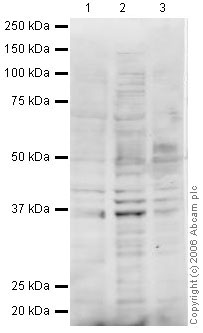
All lanes : Anti-MSY2/YBOX2 antibody (ab33164) at 1 µg/mlLane 1 : MEL-2 (Human embryonic stem cell, female cell line) Whole Cell Lysate (ab27196)Lane 2 : F9 (Mouse embryonic carcinoma cell line) Whole Cell Lysate (ab27193)Lane 3 : Embryonic Germ Cell LysateLysates/proteins at 20 µg per lane.SecondaryRabbit IgG secondary antibody (ab28446) at 1/10000 dilutionObserved band size : 37 kDa (why is the actual band size different from the predicted?)Additional bands at : 39 kDa (possible glycosylated form,post-translational modification),42 kDa (possible glycosylated form,post-translational modification).This antibody recognised a number of bands included 3 clustered around the YBOX2 predicted molecular weight of 38 kDa. We cannot be sure which of these bands, if any, corresponds to YBOX2. We are also unsure whether YBOX2 would be expected to be expressed at a detectable level for Western Blotting in the lysates tested.
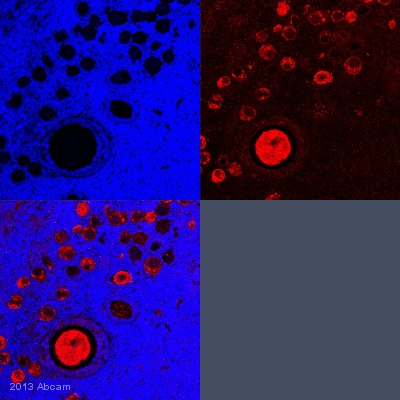
IHC-P image of MSY2/YBOX2 staining with ab33164 on tissue sections from adult marmoset ovary. The sections were subjected to heat-mediated antigen retrieval using Dako antigen retrieval solution. The sections were then blocked with 5% milk for 30 minutes at 25°C, before incubation with ab33164 (1/100 dilution) for 18 hours at 4°C. The secondary was an Alexa-Fluor 555 conjugated goat anti-rabbit polyclonal, used at a 1/500 dilution.See Abreview