Anti-MSI2 antibody [EP1305Y]
| Name | Anti-MSI2 antibody [EP1305Y] |
|---|---|
| Supplier | Abcam |
| Catalog | ab76148 |
| Prices | $391.00 |
| Sizes | 100 µl |
| Host | Rabbit |
| Clonality | Monoclonal |
| Isotype | IgG |
| Clone | EP1305Y |
| Applications | ICC/IF ICC/IF WB IHC-P FC |
| Species Reactivities | Mouse, Rat, Human |
| Antigen | A synthetic peptide derived from Human MSI2 (UniProt: Q96DH6) |
| Blocking Peptide | MSI2 peptide |
| Description | Rabbit Monoclonal |
| Gene | MSI2 |
| Conjugate | Unconjugated |
| Supplier Page | Shop |
Product images
![All lanes : Anti-MSI2 antibody [EP1305Y] (ab76148) at 1/2000 dilutionLane 1 : Rat brain cell lysates Lane 2 : Human brain cell lysates Lane 3 : SW480 cell lysates Lane 4 : T47D cell lysates Lysates/proteins at 10 µg per lane.SecondaryHRP labelled goat anti-rabbit at 1/2000 dilution](http://www.bioprodhub.com/system/product_images/ab_products/2/sub_3/25122_ab76148_1.jpg) All lanes : Anti-MSI2 antibody [EP1305Y] (ab76148) at 1/2000 dilutionLane 1 : Rat brain cell lysates Lane 2 : Human brain cell lysates Lane 3 : SW480 cell lysates Lane 4 : T47D cell lysates Lysates/proteins at 10 µg per lane.SecondaryHRP labelled goat anti-rabbit at 1/2000 dilution
All lanes : Anti-MSI2 antibody [EP1305Y] (ab76148) at 1/2000 dilutionLane 1 : Rat brain cell lysates Lane 2 : Human brain cell lysates Lane 3 : SW480 cell lysates Lane 4 : T47D cell lysates Lysates/proteins at 10 µg per lane.SecondaryHRP labelled goat anti-rabbit at 1/2000 dilution
 Immunohistochemical analysis of paraffin-embedded human placenta with ab76148 at 1/100-1/250 dilution.
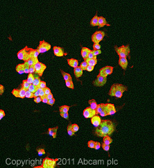 ICC/IF image of ab76148 stained PC12 cells. The cells were 4% formaldehyde fixed (10 min) and then incubated in 1%BSA / 10% normal goat serum / 0.3M glycine in 0.1% PBS-Tween for 1h to permeabilise the cells and block non-specific protein-protein interactions. The cells were then incubated with the antibody (ab76148, 1µg/ml) overnight at +4°C. The secondary antibody (green) was Alexa Fluor® 488 goat anti-rabbit IgG (H+L) used at a 1/1000 dilution for 1h. Alexa Fluor® 594 WGA was used to label plasma membranes (red) at a 1/200 dilution for 1h. DAPI was used to stain the cell nuclei (blue) at a concentration of 1.43µM.
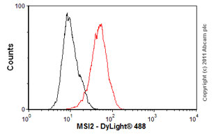 Overlay histogram showing HeLa cells stained with ab76148 (red line). The cells were fixed with 4% paraformaldehyde (10 min) and then permeabilized with 0.1% PBS-Tween for 20 min. The cells were then incubated in 1x PBS / 10% normal goat serum / 0.3M glycine to block non-specific protein-protein interactions followed by the antibody (ab76148, 1/100 dilution) for 30 min at 22ºC. The secondary antibody used was DyLight® 488 goat anti-rabbit IgG (H+L) (ab96899) at 1/500 dilution for 30 min at 22ºC. Isotype control antibody (black line) was rabbit IgG (monclonal) (1µg/1x106 cells) used under the same conditions. Acquisition of >5,000 events was performed. This antibody gave a positive result in HeLa cells fixed with 80% methanol (5 min)/permeabilized in 0.1% PBS-Tween for 20 min used under the same conditions.
Product References
Musashi2 is required for the self-renewal and pluripotency of embryonic stem - Musashi2 is required for the self-renewal and pluripotency of embryonic stem
Wuebben EL, Mallanna SK, Cox JL, Rizzino A. PLoS One. 2012;7(4):e34827.
Inactivation of a single copy of Crebbp selectively alters pre-mRNA processing in - Inactivation of a single copy of Crebbp selectively alters pre-mRNA processing in
Lemieux ME, Cheng Z, Zhou Q, White R, Cornell J, Kung AL, Rebel VI. PLoS One. 2011;6(8):e24153.
MSI2 protein expression predicts unfavorable outcome in acute myeloid leukemia. - MSI2 protein expression predicts unfavorable outcome in acute myeloid leukemia.
Byers RJ, Currie T, Tholouli E, Rodig SJ, Kutok JL. Blood. 2011 Sep 8;118(10):2857-67.