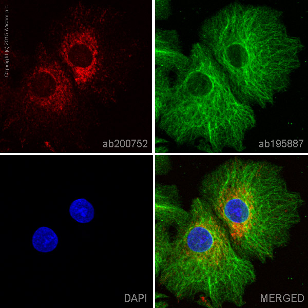
ab200752 staining Monoamine Oxidase A in HepG2 cells. The cells were fixed with 100% methanol (5 min), permeabilized with 0.1% Triton X-100 for 5 minutes and then blocked with 1% BSA/10% normal goat serum/0.3M glycine in 0.1% PBS-Tween for 1h. The cells were then incubated overnight at +4°C with ab200752 at a 1/50 dilution (shown in red) and ab195887, Mouse monoclonal to alpha Tubulin (Alexa Fluor® 488), at a 1/250 dilution (shown in green). Nuclear DNA was labelled with DAPI (shown in blue).Image was taken with a confocal microscope (Leica-Microsystems, TCS SP8).This product also gave a positive signal under the same testing conditions in HepG2 cells fixed with 4% formaldehyde (10 min).