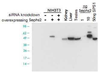
Western blot using Sephs2 polyclonal antibody ( Cat # PAB11323 ) shows detection of Sephs2 in NIH/3T3 cells over-expressing this protein. No signal is seen in control lysates or in lysates from cells over-expressing the protein after pre-treatment with Sephs2 siRNA. Endogenous Sephs2 can be detected in mouse kidney, liver and testes tissue lysates. Partial cross-reactivity is seen against recombinant SPS1. The primary antibody was used at a 1 : 1000 dilution. Personal Communication, D. Hatfield, NCI, Bethesda, MD.