Anti-MMP1 antibody [EP1247Y]
| Name | Anti-MMP1 antibody [EP1247Y] |
|---|---|
| Supplier | Abcam |
| Catalog | ab52631 |
| Prices | $392.00 |
| Sizes | 100 µl |
| Host | Rabbit |
| Clonality | Monoclonal |
| Isotype | IgG |
| Clone | EP1247Y |
| Applications | IHC-F WB ICC/IF FC IHC-P |
| Species Reactivities | Human |
| Antigen | Synthetic peptide (the amino acid sequence is considered to be commercially sensitive) corresponding to Human MMP1 aa 100-200 |
| Description | Rabbit Monoclonal |
| Gene | MMP1 |
| Conjugate | Unconjugated |
| Supplier Page | Shop |
Product images
![Anti-MMP1 antibody [EP1247Y] (ab52631) at 1/10000 dilution (unpurified) + MMP1 recombinant protein at 10 µgSecondaryPeroxidase-conjugated goat anti-rabbit IgG (H+L) at 1/1000 dilution](http://www.bioprodhub.com/system/product_images/ab_products/2/sub_3/23267_ab52631-237406-ab52631upwb.jpg) Anti-MMP1 antibody [EP1247Y] (ab52631) at 1/10000 dilution (unpurified) + MMP1 recombinant protein at 10 µgSecondaryPeroxidase-conjugated goat anti-rabbit IgG (H+L) at 1/1000 dilution
Anti-MMP1 antibody [EP1247Y] (ab52631) at 1/10000 dilution (unpurified) + MMP1 recombinant protein at 10 µgSecondaryPeroxidase-conjugated goat anti-rabbit IgG (H+L) at 1/1000 dilution
![Anti-MMP1 antibody [EP1247Y] (ab52631) at 1/10000 dilution (purified) + MMP1 recombinant protein at 10 µgSecondaryPeroxidase-conjugated goat anti-rabbit IgG (H+L) at 1/1000 dilution](http://www.bioprodhub.com/system/product_images/ab_products/2/sub_3/23268_ab52631-237405-ab52631pwb.jpg) Anti-MMP1 antibody [EP1247Y] (ab52631) at 1/10000 dilution (purified) + MMP1 recombinant protein at 10 µgSecondaryPeroxidase-conjugated goat anti-rabbit IgG (H+L) at 1/1000 dilution
Anti-MMP1 antibody [EP1247Y] (ab52631) at 1/10000 dilution (purified) + MMP1 recombinant protein at 10 µgSecondaryPeroxidase-conjugated goat anti-rabbit IgG (H+L) at 1/1000 dilution
![Anti-MMP1 antibody [EP1247Y] (ab52631) at 1/1000 dilution (unpurified) + SKBr-3 cell lysate at 10 µgSecondaryGoat anti-rabbit HRP at 1/2000 dilution](http://www.bioprodhub.com/system/product_images/ab_products/2/sub_3/23269_mmp11.jpg) Anti-MMP1 antibody [EP1247Y] (ab52631) at 1/1000 dilution (unpurified) + SKBr-3 cell lysate at 10 µgSecondaryGoat anti-rabbit HRP at 1/2000 dilution
Anti-MMP1 antibody [EP1247Y] (ab52631) at 1/1000 dilution (unpurified) + SKBr-3 cell lysate at 10 µgSecondaryGoat anti-rabbit HRP at 1/2000 dilution
![All lanes : Anti-MMP1 antibody [EP1247Y] (ab52631) at 1/1000 dilution (unpurified)Lane 1 : Human colon mucosa tissue lysate (inflamed)Lane 2 : Human colon mucosa tissue lysate (uninflamed)Lysates/proteins at 40 µg per lane.SecondaryHRP-conjugated Goat anti-rabbit IgG polyclonal at 1/3000 dilutiondeveloped using the ECL techniquePerformed under reducing conditions.](http://www.bioprodhub.com/system/product_images/ab_products/2/sub_3/23270_MMP1-Primary-antibodies-ab52631-12.jpg) All lanes : Anti-MMP1 antibody [EP1247Y] (ab52631) at 1/1000 dilution (unpurified)Lane 1 : Human colon mucosa tissue lysate (inflamed)Lane 2 : Human colon mucosa tissue lysate (uninflamed)Lysates/proteins at 40 µg per lane.SecondaryHRP-conjugated Goat anti-rabbit IgG polyclonal at 1/3000 dilutiondeveloped using the ECL techniquePerformed under reducing conditions.
All lanes : Anti-MMP1 antibody [EP1247Y] (ab52631) at 1/1000 dilution (unpurified)Lane 1 : Human colon mucosa tissue lysate (inflamed)Lane 2 : Human colon mucosa tissue lysate (uninflamed)Lysates/proteins at 40 µg per lane.SecondaryHRP-conjugated Goat anti-rabbit IgG polyclonal at 1/3000 dilutiondeveloped using the ECL techniquePerformed under reducing conditions.
 Immunohistochemistry (Formalin/PFA-fixed paraffin-embedded sections) analysis of human testis tissue labelling MMP1 with unpurified ab52631 at 1/60. Heat mediated antigen retrieval was performed using Tris/EDTA buffer pH 9. A prediluted HRP-polymer conjugated anti-rabbit IgG was used as the secondary antibody. Negative control using PBS instead of primary antibody. Counterstained with hematoxylin.
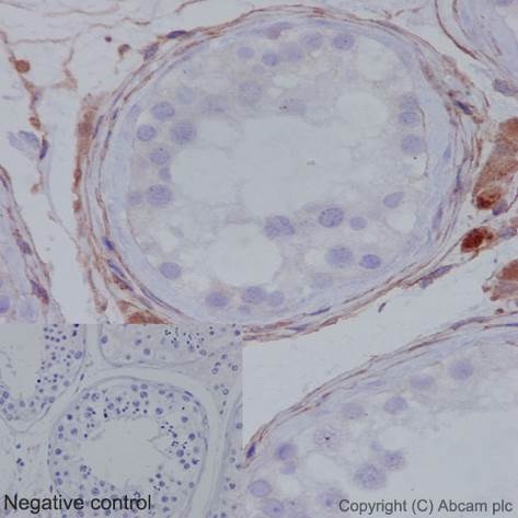 Immunohistochemistry (Formalin/PFA-fixed paraffin-embedded sections) analysis of human testis tissue labelling MMP1 with purified ab52631 at 1/100. Heat mediated antigen retrieval was performed using Tris/EDTA buffer pH 9. A prediluted HRP-polymer conjugated anti-rabbit IgG was used as the secondary antibody. Negative control using PBS instead of primary antibody. Counterstained with hematoxylin.
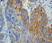 Immunohistochemistry (Formalin/PFA-fixed paraffin-embedded sections) analysis of human cervical carcinoma tissue labelling MMP1 with ab52631 at 1/50.
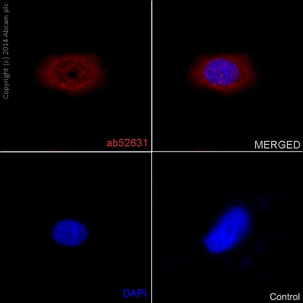 Immunocytochemistry/Immunofluorescence analysis of HeLa cells labelling MMP1 with unpurified ab52631 at 1/30. Cells were fixed with 4% paraformaldehyde. An Alexa Fluor® 555-conjugated goat anti-rabbit IgG (1/500) was used as the secondary antibody. DAPI (blue) was used as the nuclear counterstain.Control: primary antibody (1/30) and secondary antibody, ab150120, an Alexa Fluor® 594-conjugated goat anti-mouse IgG (1/500).
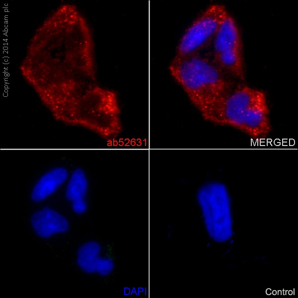 Immunocytochemistry/Immunofluorescence analysis of HeLa cells labelling MMP1 with purified ab52631 at 1/50. Cells were fixed with 4% paraformaldehyde. An Alexa Fluor® 555-conjugated goat anti-rabbit IgG (1/500) was used as the secondary antibody. DAPI (blue) was used as the nuclear counterstain.Control: primary antibody (1/50) and secondary antibody, ab150120, an Alexa Fluor® 594-conjugated goat anti-mouse IgG (1/500).
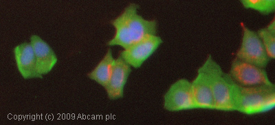 ICC/IF image of unpurified ab52631 stained MCF7 cells. The cells were 4% PFA fixed (10 min) and then incubated in 1% BSA / 10% normal goat serum / 0.3M glycine in 0.1% PBS-Tween for 1h to permeabilise the cells and block non-specific protein-protein interactions. The cells were then incubated with the antibody (ab52631, 1/1000 dilution) overnight at +4°C. The secondary antibody (green) was Alexa Fluor® 488 goat anti-rabbit IgG (H+L) used at a 1/1000 dilution for 1h. Alexa Fluor® 594 WGA was used to label plasma membranes (red) at a 1/200 dilution for 1h. DAPI was used to stain the cell nuclei (blue) at a concentration of 1.43µM.
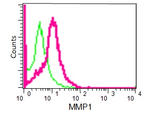 Flow cytometry analysis of HeLa cells labelling MMP1 with unpurified ab52631 at 1/50 (red). Cells were fixed with 2% paraformaldehyde. A FITC-conjugated goat anti-rabbit IgG (1/150) was used as the secondary antibody. Green - Isotype control, rabbit monoclonal IgG.
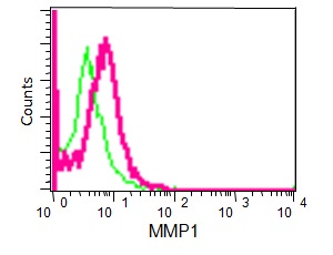 Flow cytometry analysis of HeLa cells labelling MMP1 with purified ab52631 at 1/70 (red). Cells were fixed with 2% paraformaldehyde. A FITC-conjugated goat anti-rabbit IgG was used as the secondary antibody (1/150). Green - Isotype control, rabbit monoclonal IgG.
Product References
Structural modifications and tissue response after standard epi-off and - Structural modifications and tissue response after standard epi-off and
Mastropasqua L, Lanzini M, Curcio C, Calienno R, Mastropasqua R, Colasante M, Mastropasqua A, Nubile M. Invest Ophthalmol Vis Sci. 2014 Apr 17;55(4):2526-33.
Calcipotriol counteracts betamethasone-induced decrease in extracellular matrix - Calcipotriol counteracts betamethasone-induced decrease in extracellular matrix
Norsgaard H, Kurdykowski S, Descargues P, Gonzalez T, Marstrand T, Dunstl G, Ropke M. Arch Dermatol Res. 2014 Oct;306(8):719-29.
The effects of retinoic acid on human corneal stromal keratocytes cultured in - The effects of retinoic acid on human corneal stromal keratocytes cultured in
Gouveia RM, Connon CJ. Invest Ophthalmol Vis Sci. 2013 Nov 13;54(12):7483-91. doi:
Differential expression of degradome components in cutaneous squamous cell - Differential expression of degradome components in cutaneous squamous cell
Prasad NB, Fischer AC, Chuang AY, Wright JM, Yang T, Tsai HL, Westra WH, Liegeois NJ, Hess AD, Tufaro AP. Mod Pathol. 2014 Jul;27(7):945-57.
Inactivation of Rb in stromal fibroblasts promotes epithelial cell invasion. - Inactivation of Rb in stromal fibroblasts promotes epithelial cell invasion.
Pickard A, Cichon AC, Barry A, Kieran D, Patel D, Hamilton P, Salto-Tellez M, James J, McCance DJ. EMBO J. 2012 May 29;31(14):3092-103.
The role of MMP-1 in breast cancer growth and metastasis to the brain in a - The role of MMP-1 in breast cancer growth and metastasis to the brain in a
Liu H, Kato Y, Erzinger SA, Kiriakova GM, Qian Y, Palmieri D, Steeg PS, Price JE. BMC Cancer. 2012 Dec 7;12:583.
Comprehensive analysis of leukocytes, vascularization and matrix - Comprehensive analysis of leukocytes, vascularization and matrix
Guo Y, He B, Xu X, Wang J. PLoS One. 2011 Feb 17;6(2):e16840.
Expression of osteonectin/secreted protein acidic and rich in cysteine and matrix - Expression of osteonectin/secreted protein acidic and rich in cysteine and matrix
Shen LC, Chen YK, Hsue SS, Shaw SY. J Oral Pathol Med. 2010 Mar;39(3):242-9.
Cell-specific gene expression in Langerhans cell histiocytosis lesions reveals a - Cell-specific gene expression in Langerhans cell histiocytosis lesions reveals a
Allen CE, Li L, Peters TL, Leung HC, Yu A, Man TK, Gurusiddappa S, Phillips MT, Hicks MJ, Gaikwad A, Merad M, McClain KL. J Immunol. 2010 Apr 15;184(8):4557-67.