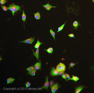
ab75506 stained U-87 MG cells. The cells were 4% formaldehyde fixed (10 min) and then incubated in 1%BSA / 10% normal goat serum / 0.3M glycine in 0.1% PBS-Tween for 1h to permeabilise the cells and block non-specific protein-protein interactions. The cells were then incubated with the antibody ab75506 at 5µg/ml overnight at +4°C. The secondary antibody (green) was DyLight® 488 goat anti- rabbit (ab96899) IgG (H+L) used at a 1/250 dilution for 1h. Alexa Fluor® 594 WGA was used to label plasma membranes (red) at a 1/200 dilution for 1h. DAPI was used to stain the cell nuclei (blue) at a concentration of 1.43µM.
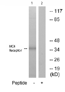
All lanes : Anti-MC4 Receptor antibody (ab75506) at 1/500 dilutionLane 1 : Extracts from MCF-7 cellsLane 2 : Extracts from MCF-7 cells plus 5µg immunizing peptideLysates/proteins at 5 µg per lane.

IHC-P image of MC4 Receptor staining on human placenta sections using ab75506 (1:1000). The sections were de-paraffinized and subjected to heat mediated antigen retrieval using Citric acid pH6. The sections were blocked using 1% BSA for 10 mins at 21°C. Primary antibody was incubated at 21°C for 2 hours.See Abreview