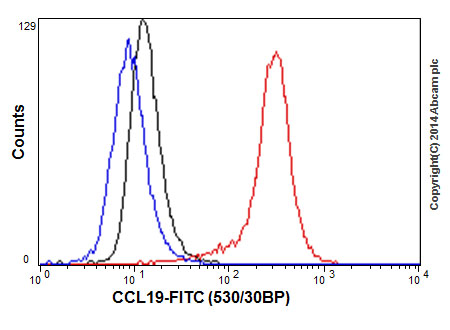
Flow cytometric analysis of 2% paraformaldehyde-fixed A549 cells labeling Macrophage Inflammatory Protein 3 beta with ab192877 at 1/170 dilution (red) compared with a rabbit monoclonal IgG isotype control (black) and an unlabelled control (cells without incubation with primary antibody and secondary antibody; blue). Goat anti rabbit IgG (FITC) at 1/150 dilution was used as the secondary antibody.
![Anti-Macrophage Inflammatory Protein 3 beta antibody [EPR7044(2)] (ab192877) at 1/20000 dilution + Human Macrophage Inflammatory Protein 3 beta recombinant protein (aa22-98) at 10 µgSecondaryGoat Anti-Rabbit IgG, (H+L), Peroxidase conjugate at 1/1000 dilution](http://www.bioprodhub.com/system/product_images/ab_products/2/sub_3/18448_ab192877-230668-ab192877WBa.jpg)
Anti-Macrophage Inflammatory Protein 3 beta antibody [EPR7044(2)] (ab192877) at 1/20000 dilution + Human Macrophage Inflammatory Protein 3 beta recombinant protein (aa22-98) at 10 µgSecondaryGoat Anti-Rabbit IgG, (H+L), Peroxidase conjugate at 1/1000 dilution

![Anti-Macrophage Inflammatory Protein 3 beta antibody [EPR7044(2)] (ab192877) at 1/20000 dilution + Human Macrophage Inflammatory Protein 3 beta recombinant protein (aa22-98) at 10 µgSecondaryGoat Anti-Rabbit IgG, (H+L), Peroxidase conjugate at 1/1000 dilution](http://www.bioprodhub.com/system/product_images/ab_products/2/sub_3/18448_ab192877-230668-ab192877WBa.jpg)