Anti-Ly6g antibody [RB6-8C5] (Allophycocyanin)
| Name | Anti-Ly6g antibody [RB6-8C5] (Allophycocyanin) |
|---|---|
| Supplier | Abcam |
| Catalog | ab25273 |
| Prices | $392.00 |
| Sizes | 50 µg |
| Host | Rat |
| Clonality | Monoclonal |
| Isotype | IgG2b |
| Clone | RB6-8C5 |
| Applications | FC IHC-F IP WB |
| Species Reactivities | Mouse, Human, Monkey |
| Antigen | The details of the immunogen for this antibody are not available |
| Description | Rat Monoclonal |
| Gene | Ly6g |
| Conjugate | Allophycocyanin |
| Supplier Page | Shop |
Product images
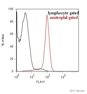 ab25273 at a 1/100 dilution detecting Ly6g in human buffy coat cells by Flow Cytometry. See Abreview
 ab25273 at a 1/100 dilution detecting Ly6g (Gr1) in murine blood cells by Flow Cytometry.See Abreview
 ab25273 staining Ly6g in Mouse spleen tissue sections by Immunohistochemistry (IHC-Fr - frozen sections). Tissue was fixed with acetone and blocked with 5% Goat serum for 1 hour at 25°C. Samples were incubated with primary antibody (1/100 in 5% Goat serum) for 1 hour at 4°C. An Alexa Fluor® 488-conjugated Goat anti-rat IgG polyclonal (1/400) was used as the secondary antibody.See Abreview