Anti-LXR beta antibody
| Name | Anti-LXR beta antibody |
|---|---|
| Supplier | Abcam |
| Catalog | ab28479 |
| Prices | $376.00 |
| Sizes | 100 µg |
| Host | Rabbit |
| Clonality | Polyclonal |
| Isotype | IgG |
| Applications | WB ICC/IF ICC/IF |
| Species Reactivities | Mouse, Rat, Human |
| Antigen | Synthetic peptide: RYACRGSG TCQMDA , corresponding to amino acids 113-126 of Rat LXR beta |
| Description | Rabbit Polyclonal |
| Gene | NR1H2 |
| Conjugate | Unconjugated |
| Supplier Page | Shop |
Product images
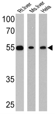 All lanes : Anti-LXR beta antibody (ab28479) at 1/1000 dilutionLane 1 : Rat liver cell lysateLane 2 : Mouse liver cell lysateLane 3 : HeLa cell lysateLysates/proteins at 25 µg per lane.SecondaryHRP-conjugated anti-rabbit
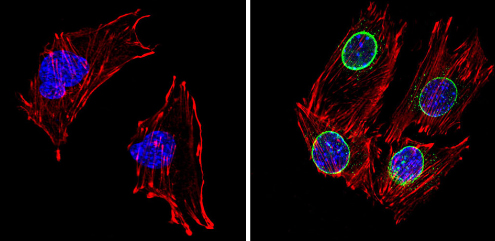 ab28479 labelling LXR beta (green) in the nuclear envelope of C2C12 cells (right) compared with a negative control (left) by Immunocytochemistry/Immunofluorescence. Formalin-fixed cells were permeabilized with 0.1% Triton X-100 in TBS for 5-10 minutes, and blocked with 3% BSA-PBS for 30 minutes at room temperature. Cells were incubated with the primary antibody (1:200 in 3% BSA-PBS) overnight at 4 ºC. A DyLight 488-conjugated Goat anti-rabbit IgG was used as the secondary antibody. Red (phalloidin) - F-actin, Blue (DAPI) - nuclei. Images were taken at a magnification of 60x.