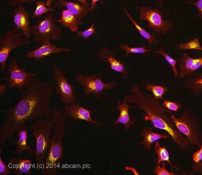
ICC/IF image of ab125006 stained HeLa cells. The cells were 100% methanol fixed (5 min) and then incubated in 1%BSA / 10% normal goat serum / 0.3M glycine in 0.1% PBS-Tween for 1h to permeabilise the cells and block non-specific protein-protein interactions. The cells were then incubated with the antibody ab125006 at 1/100 dilution overnight at +4°C. The secondary antibody (pseudo-colored green) was Alexa Fluor® 488 goat anti- rabbit (ab150081) IgG (H+L) preadsorbed, used at a 1/1000 dilution for 1h. Alexa Fluor® 594 WGA was used to label plasma membranes (pseudo-colored red) at a 1/200 dilution for 1h at room temperature. DAPI was used to stain the cell nuclei (pseudo-colored blue) at a concentration of 1.43µM for 1hour at room temperature.
![All lanes : Anti-LMAN1 antibody [EPR6979] (ab125006) at 1/1000 dilutionLane 1 : HeLa cell lysateLane 2 : 293T cell lysateLane 3 : SH-SY5Y cell lysateLane 4 : NCI-H460 cell lysateLane 5 : Jurkat cell lysateLane 6 : JAR cell lysateLysates/proteins at 10 µg per lane.SecondaryGoat anti-Rabbit HRP at 1/2000 dilution](http://www.bioprodhub.com/system/product_images/ab_products/2/sub_3/16706_LMAN1-Primary-antibodies-ab125006-1.JPG)
All lanes : Anti-LMAN1 antibody [EPR6979] (ab125006) at 1/1000 dilutionLane 1 : HeLa cell lysateLane 2 : 293T cell lysateLane 3 : SH-SY5Y cell lysateLane 4 : NCI-H460 cell lysateLane 5 : Jurkat cell lysateLane 6 : JAR cell lysateLysates/proteins at 10 µg per lane.SecondaryGoat anti-Rabbit HRP at 1/2000 dilution
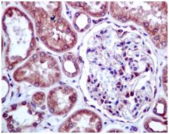
ab125006 at 1/100 dilution staining LMAN1 in paraffin-embedded Human kidney tissue by Immunohistochemistry.
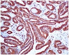
ab125006 at 1/100 dilution staining LMAN1 in paraffin-embedded Human papillary carcinoma of thyroid gland tissue by Immunohistochemistry.