Anti-liver FABP antibody
| Name | Anti-liver FABP antibody |
|---|---|
| Supplier | Abcam |
| Catalog | ab7847 |
| Prices | $400.00 |
| Sizes | 500 µl |
| Host | Rabbit |
| Clonality | Polyclonal |
| Isotype | IgG |
| Applications | ICC/IF ICC/IF IP WB |
| Species Reactivities | Mouse, Rat, Human, Pig |
| Description | Rabbit Polyclonal |
| Gene | FABP1 |
| Conjugate | Unconjugated |
| Supplier Page | Shop |
Product images
![liver FABP was immunoprecipitated using 0.5mg Mouse Liver tissue extract, 5µg of Rabbit polyclonal to liver FABP and 50µl of protein G magnetic beads (+). No antibody was added to the control (-).The antibody was incubated under agitation with Protein G beads for 10min, Mouse Liver tissue extract lysate diluted in RIPA buffer was added to each sample and incubated for a further 10min under agitation.Proteins were eluted by addition of 40µl SDS loading buffer and incubated for 10min at 70°C; 10µl of each sample was separated on a SDS PAGE gel, transferred to a nitrocellulose membrane, blocked with 5% BSA and probed with ab7847.Secondary: Mouse monoclonal [SB62a] Secondary Antibody to Rabbit IgG light chain (HRP) (ab99697).Band: 13kDa; liver FABP](http://www.bioprodhub.com/system/product_images/ab_products/2/sub_3/16610_ab7847-199602-IPV024ab784720m.jpg) liver FABP was immunoprecipitated using 0.5mg Mouse Liver tissue extract, 5µg of Rabbit polyclonal to liver FABP and 50µl of protein G magnetic beads (+). No antibody was added to the control (-).The antibody was incubated under agitation with Protein G beads for 10min, Mouse Liver tissue extract lysate diluted in RIPA buffer was added to each sample and incubated for a further 10min under agitation.Proteins were eluted by addition of 40µl SDS loading buffer and incubated for 10min at 70°C; 10µl of each sample was separated on a SDS PAGE gel, transferred to a nitrocellulose membrane, blocked with 5% BSA and probed with ab7847.Secondary: Mouse monoclonal [SB62a] Secondary Antibody to Rabbit IgG light chain (HRP) (ab99697).Band: 13kDa; liver FABP
liver FABP was immunoprecipitated using 0.5mg Mouse Liver tissue extract, 5µg of Rabbit polyclonal to liver FABP and 50µl of protein G magnetic beads (+). No antibody was added to the control (-).The antibody was incubated under agitation with Protein G beads for 10min, Mouse Liver tissue extract lysate diluted in RIPA buffer was added to each sample and incubated for a further 10min under agitation.Proteins were eluted by addition of 40µl SDS loading buffer and incubated for 10min at 70°C; 10µl of each sample was separated on a SDS PAGE gel, transferred to a nitrocellulose membrane, blocked with 5% BSA and probed with ab7847.Secondary: Mouse monoclonal [SB62a] Secondary Antibody to Rabbit IgG light chain (HRP) (ab99697).Band: 13kDa; liver FABP
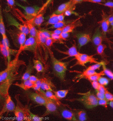 ICC/IF image of ab7847 stained HepG2 cells. The cells were 4% PFA fixed (10 min) and then incubated in 1%BSA / 10% normal goat serum / 0.3M glycine in 0.1% PBS-Tween for 1h to permeabilise the cells and block non-specific protein-protein interactions. The cells were then incubated with the antibody (ab7847, 5µg/ml) overnight at +4°C. The secondary antibody (green) was Alexa Fluor® 488 goat anti-rabbit IgG (H+L) used at a 1/1000 dilution for 1h. Alexa Fluor® 594 WGA was used to label plasma membranes (red) at a 1/200 dilution for 1h. DAPI was used to stain the cell nuclei (blue) at a concentration of 1.43µM.
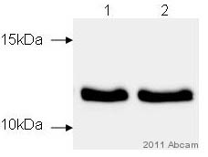 All lanes : Anti-liver FABP antibody (ab7847) at 1/1000 dilutionLane 1 : Whole tissue lysate prepared from pig liverLane 2 : Whole tissue lysate prepared from pig liverLysates/proteins at 10 µg per lane.SecondaryGoat anti-rabbit HRP at 1/2000 dilutionImage courtesy of an anonymous Abreview.
Product References
Characterization of 4-HNE modified L-FABP reveals alterations in structural and - Characterization of 4-HNE modified L-FABP reveals alterations in structural and
Smathers RL, Fritz KS, Galligan JJ, Shearn CT, Reigan P, Marks MJ, Petersen DR. PLoS One. 2012;7(6):e38459.
Ablation of PI3K p110-alpha prevents high-fat diet-induced liver steatosis. - Ablation of PI3K p110-alpha prevents high-fat diet-induced liver steatosis.
Chattopadhyay M, Selinger ES, Ballou LM, Lin RZ. Diabetes. 2011 May;60(5):1483-92.
Thyroid hormone (T3) and TRbeta agonist GC-1 inhibit/reverse nonalcoholic fatty - Thyroid hormone (T3) and TRbeta agonist GC-1 inhibit/reverse nonalcoholic fatty
Perra A, Simbula G, Simbula M, Pibiri M, Kowalik MA, Sulas P, Cocco MT, Ledda-Columbano GM, Columbano A. FASEB J. 2008 Aug;22(8):2981-9.
An improved method for the purification of rat liver-type fatty acid binding - An improved method for the purification of rat liver-type fatty acid binding
Velkov T, Chuang S, Prankerd R, Sakellaris H, Porter CJ, Scanlon MJ. Protein Expr Purif. 2005 Nov;44(1):23-31.