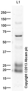
Anti-Lipocalin 2 antibody (ab41105) at 1 µg/ml + Colon tissue lysate (Human) Tissue Lysate - adult normal tissue (ab30051) at 30 µgSecondaryGoat Anti-Rabbit IgG H&L (HRP) (ab97051) at 1/10000 dilutiondeveloped using the ECL techniquePerformed under reducing conditions.
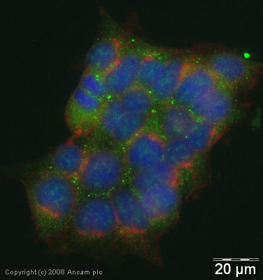
ICC/IF image of ab41105 stained human Hek293 cells. The cells were methanol fixed (10 min), permabilised in 0.1% PBS-Tween (20 min) and incubated with the antibody (ab41105, 5µg/ml) for 1h at room temperature. 1%BSA / 10% normal goat serum / 0.3M glycine was used to block non-specific protein-protein interactions. The secondary antibody (green) was Alexa Fluor® 488 goat anti-rabbit IgG (H+L) used at a 1/1000 dilution for 1h. Alexa Fluor® 594 WGA was used to label plasma membranes (red). DAPI was used to stain the cell nuclei (blue).

ab41105 staining Lipocalin 2 in human lung tissue by Immunohistochemistry (Formalin/PFA-fixed paraffin embedded sections). Tissue underwent fixation in formaldehyde, heat mediated antigen retrieval in Citrate buffer pH6.0 and blocking for 15 minutes at 20°C (5 minutes peroxidase block and 10 minutes protein blocks). The primary antibody was diluted 1/250 or 1/1000 and incubated with sample for 45 minutes at 20°C. A HRP-conjugated goat polyclonal to rabbit IgG was used undiluted as the secondary. See Abreview

ab41105 staining Lipocalin 2 in rat lung tissue section by Immunohistochemistry (Formalin/PFA-fixed paraffin embedded sections). Tissue underwent fixation in formaldehyde, heat mediated antigen retrieval in Citrate buffer pH6.0 and blocking for 15 minutes at 20°C (5 minutes peroxidase block and 10 minutes protein blocks). The primary antibody was diluted 1/250 or 1/1000 and incubated with sample for 45 minutes at 20°C. A HRP-conjugated goat polyclonal to rabbit IgG was used undiluted as the secondary. See Abreview