Anti-LIPG antibody
| Name | Anti-LIPG antibody |
|---|---|
| Supplier | Abcam |
| Catalog | ab100987 |
| Prices | $370.00 |
| Sizes | 200 µl |
| Host | Rabbit |
| Clonality | Polyclonal |
| Isotype | IgG |
| Applications | WB ICC/IF ICC/IF |
| Species Reactivities | Human, Rabbit, Bovine, Pig |
| Antigen | Synthetic peptide conjugated to KLH derived from within residues 350 - 450 of Human LIPG |
| Description | Rabbit Polyclonal |
| Gene | LIPG |
| Conjugate | Unconjugated |
| Supplier Page | Shop |
Product images
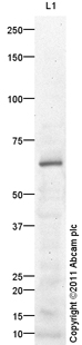 Anti-LIPG antibody (ab100987) at 1/250 dilution + Liver (Human) Tissue Lysate - adult normal tissue (ab29889) at 10 µgSecondaryGoat Anti-Rabbit IgG H&L (HRP) preadsorbed (ab97080) at 1/5000 dilutiondeveloped using the ECL techniquePerformed under reducing conditions.
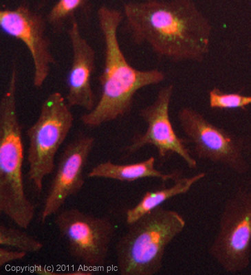 ICC/IF image of ab100987 stained HeLa cells. The cells were 4% PFA fixed (10 min) and then incubated in 1%BSA / 10% normal goat serum / 0.3M glycine in 0.1% PBS-Tween for 1h to permeabilise the cells and block non-specific protein-protein interactions. The cells were then incubated with the antibody (ab100987, µg/ml) overnight at +4°C. The secondary antibody (green) was ab96899, DyLight® 488 goat anti-rabbit IgG (H+L) used at a 1/250 dilution for 1h. Alexa Fluor® 594 WGA was used to label plasma membranes (red) at a 1/200 dilution for 1h. DAPI was used to stain the cell nuclei (blue) at a concentration of 1.43µM. This antibody also gave a positive result in 100% methanol fixed (5 min) HeLa cells at 5µg/ml.