![All lanes : Anti-Keap1 antibody [EPR1805(2)] (ab181144) at 1/1000 dilutionLane 1 : HeLa cell lysateLane 2 : Raji cell lysateLane 3 : NIH:OVCAR-3 cell lysateLane 4 : 293 cell lysateLysates/proteins at 10 µg per lane.SecondaryGoat Anti-Rabbit IgG, (H+L), Peroxidase conjugate at 1/1000 dilution](http://www.bioprodhub.com/system/product_images/ab_products/2/sub_3/12345_ab181144-215047-ab181144.jpg)
All lanes : Anti-Keap1 antibody [EPR1805(2)] (ab181144) at 1/1000 dilutionLane 1 : HeLa cell lysateLane 2 : Raji cell lysateLane 3 : NIH:OVCAR-3 cell lysateLane 4 : 293 cell lysateLysates/proteins at 10 µg per lane.SecondaryGoat Anti-Rabbit IgG, (H+L), Peroxidase conjugate at 1/1000 dilution
Immunohistochemical analysis of paraffin-embedded Human skeletal muscle tissue labeling Keap1 with ab181144 at 1/100 dilution followed by pre-diluted HRP-conjugated secondary antibody and counter-stained with Hematoxylin.
Immunohistochemical analysis of paraffin-embedded Human prostate tissue labeling Keap1 with ab181144 at 1/100 dilution followed by pre-diluted HRP-conjugated secondary antibody and counter-stained with Hematoxylin.
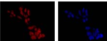
Immunofluorescent analysis of 293 cells (paraformaldehyde-fixed, 4%) labeling Keap1 with ab181144 at 1/250 dilution followed by Goat anti rabbit IgG (Alexa Fluor® 555) secondary at 1/200 dilution and counter-stained with DAPI (blue).
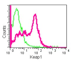
Flow cytometric analysis of Raji cells (paraformaldehyde-fixed, 2%) labeling Keap1 with ab181144 at 1/390 dilution (red) or a rabbit IgG (negative) (green), followed by Goat anti rabbit IgG (FITC) secondary at 1/150 dilution.