![All lanes : Anti-Keap1 antibody [1B4] (ab119403) at 1/500 dilutionLane 1 : HEK293T cell lysate transfected with pCMV6-ENTRY control cDNALane 2 : HEK293T cell lysate transfected with pCMV6-ENTRY Keap1 cDNALysates/proteins at 5 µg per lane.developed using the ECL technique](http://www.bioprodhub.com/system/product_images/ab_products/2/sub_3/12333_Keap1-Primary-antibodies-ab119403-1.jpg)
All lanes : Anti-Keap1 antibody [1B4] (ab119403) at 1/500 dilutionLane 1 : HEK293T cell lysate transfected with pCMV6-ENTRY control cDNALane 2 : HEK293T cell lysate transfected with pCMV6-ENTRY Keap1 cDNALysates/proteins at 5 µg per lane.developed using the ECL technique
![All lanes : Anti-Keap1 antibody [1B4] (ab119403) at 1/500 dilutionLane 1 : HepG2 cell extractLane 2 : HeLa cell extractLane 3 : SVT2 cell extractLane 4 : A549 cell extractLane 5 : COS7 cell extractLane 6 : Jurkat cell extractLane 7 : MDCK cell extractLane 8 : PC12 cell extractLane 9 : MCF7 cell extractLysates/proteins at 35 µg per lane.developed using the ECL technique](http://www.bioprodhub.com/system/product_images/ab_products/2/sub_3/12334_Keap1-Primary-antibodies-ab119403-2.JPG)
All lanes : Anti-Keap1 antibody [1B4] (ab119403) at 1/500 dilutionLane 1 : HepG2 cell extractLane 2 : HeLa cell extractLane 3 : SVT2 cell extractLane 4 : A549 cell extractLane 5 : COS7 cell extractLane 6 : Jurkat cell extractLane 7 : MDCK cell extractLane 8 : PC12 cell extractLane 9 : MCF7 cell extractLysates/proteins at 35 µg per lane.developed using the ECL technique
ab119403 at 1/150 dilution staining Keap1 in paraffin-embedded Human endometrium tissue by Immunohistochemistry.
ab119403 at 1/150 dilution staining Keap1 in paraffin-embedded Human endometrium adenocarcinoma tissue by Immunohistochemistry.
ab119403 at 1/150 dilution staining Keap1 in paraffin-embedded Human bladder carcinoma tissue by Immunohistochemistry.
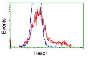
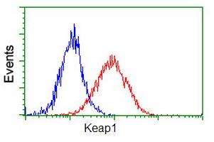
ab119403 at 1/100 dilution staining Keap1 in HEK293T cells transfected with either pCMV6-ENTRY Keap1 overexpress plasmid (Red) or empty vector control plasmid (Blue) and then analysed by Flow Cytometry.
ab119403 at 1/100 dilution staining Keap1 in HeLa cells by Flow cytometry (Red) compared to a nonspecific negative control antibody (Blue).
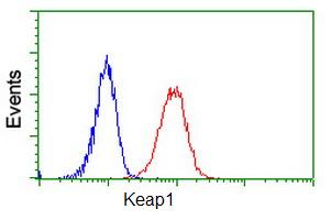
ab119403 at 1/100 dilution staining Keap1 in Jurkat cells by Flow cytometry (Red) compared to a nonspecific negative control antibody (Blue).