Anti-IQGAP1 antibody
| Name | Anti-IQGAP1 antibody |
|---|---|
| Supplier | Abcam |
| Catalog | ab86064 |
| Prices | $394.00 |
| Sizes | 100 µl |
| Host | Rabbit |
| Clonality | Polyclonal |
| Isotype | IgG |
| Applications | ICC/IF ICC/IF WB IP IHC-P IHC-F |
| Species Reactivities | Human, Dog, Chimpanzee, Ferret, Monkey, Ape, Elephant |
| Antigen | Synthetic peptide, corresponding to a region between residues 1 and 50 of human IQGAP1 (NP_003861 |
| Description | Rabbit Polyclonal |
| Gene | IQGAP1 |
| Conjugate | Unconjugated |
| Supplier Page | Shop |
Product images
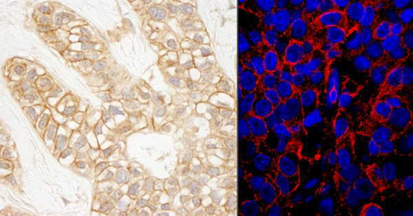 Immunohistochemistry (Formalin/PFA-fixed paraffin-embedded sections) analysis of human breast carcinoma tissue labelling IQGAP1 with ab86064 at 1/1000 (0.2µg/ml) and 1/200 (1µg/ml). Detection: DAB and a DyLight® 594-conjugated goat anti-rabbit IgG (H+L) (1/100).
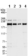 All lanes : Anti-IQGAP1 antibody (ab86064) at 0.04 µg/mlLane 1 : HeLa whole cell lysate at 50 µgLane 2 : HeLa whole cell lysate at 15 µgLane 3 : HeLa whole cell lysate at 5 µgLane 4 : 293T whole cell lysate at 50 µg
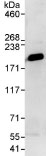 Detection of IQGAP1 by Western Blot of Immunprecipitate. ab86064 at 0.4µg/ml staining IQGAP1 in HeLa whole cell lysate immunoprecipitated using ab86064 at 3µg/mg lysate (1 mg/IP; 20% of IP loaded/lane). Detection: Chemiluminescence with exposure time of 10 seconds.
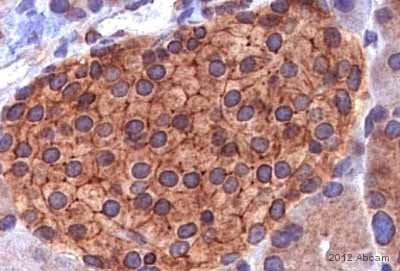 ab86064 staining IQGAP1 in human pacreatic islets by Immunohistochemistry (Formalin/PFA-fixed paraffin-embedded sections).Tissue was fixed in paraformaldehyde and a heat mediated antigen retrieval step was performed using citrate. Samples were then blocked with 5% serum for 1 hour at 20°C and then incubated with ab86064 at a 1/100 dilution for 8 hours at 4°C. The secondary used was a biotin conjugated goat anti-rabbit polyclonal used at a 1/1000 dilution.See Abreview
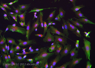 ab86064 stained SKNSH cells. The cells were 4% formaldehyde fixed (10 min) and then incubated in 1%BSA / 10% normal goat serum / 0.3M glycine in 0.1% PBS-Tween for 1h to permeabilise the cells and block non-specific protein-protein interactions. The cells were then incubated with the antibody ab86064 at 1µg/ml overnight at +4°C. The secondary antibody (green) was DyLight® 488 goat anti- rabbit (ab96899) IgG (H+L) used at a 1/1000 dilution for 1h. Alexa Fluor® 594 WGA was used to label plasma membranes (red) at a 1/200 dilution for 1h. DAPI was used to stain the cell nuclei (blue) at a concentration of 1.43µM. This antibody also gave a positive result in Methanol fixed (100%, 5min) SKNSH cells at 1ug/ml.
Product References
IQGAP1 scaffold-kinase interaction blockade selectively targets RAS-MAP - IQGAP1 scaffold-kinase interaction blockade selectively targets RAS-MAP
Jameson KL, Mazur PK, Zehnder AM, Zhang J, Zarnegar B, Sage J, Khavari PA. Nat Med. 2013 May;19(5):626-30.
A conserved role of IQGAP1 in regulating TOR complex 1. - A conserved role of IQGAP1 in regulating TOR complex 1.
Tekletsadik YK, Sonn R, Osman MA. J Cell Sci. 2012 Apr 15;125(Pt 8):2041-52.