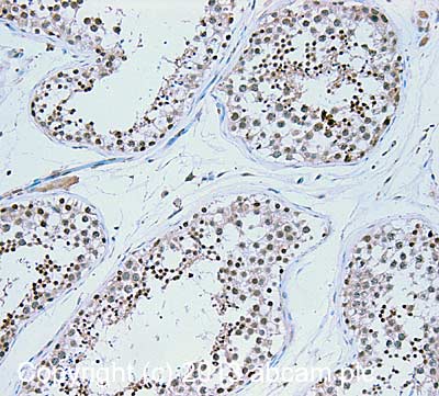
IHC image of IPL-1/STK13/Aurora C staining in Human normal testis formalin fixed paraffin embedded tissue section, performed on a Leica Bond™ system using the standard protocol F. The section was pre-treated using heat mediated antigen retrieval with sodium citrate buffer (pH6, epitope retrieval solution 1) for 20 mins. The section was then incubated with ab38299, 5µg/ml, for 15 mins at room temperature and detected using an HRP conjugated compact polymer system. DAB was used as the chromogen. The section was then counterstained with haematoxylin and mounted with DPX. For other IHC staining systems (automated and non-automated) customers should optimize variable parameters such as antigen retrieval conditions, primary antibody concentration and antibody incubation times.
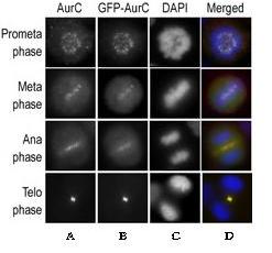
Immunofluorescence assay using HeLa cells expressing GFP-Aurora C was performed at different cellular mitotic stages, using ab38299 as primary antibody (A), GFP fluorescence (B), DAPI nuclear staining (C), and ab38299 merged to DAPI staining (column D).
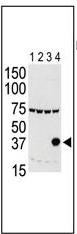
All lanes : Anti-IPL-1/STK13/Aurora C antibody (ab38299) at 1/500 dilutionLane 1 : 293 cell lysate expressing Flag tagLane 2 : 293 cell lysate expressing Flag tag Aurora A Lane 3 : 293 cell lysate expressing Flag tag Aurora BLane 4 : 293 cell lysate expressing Flag tag Aurora C