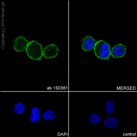
Immunofluorescent analysis of 4% paraformaldehyde-fixed, 0.1% Triton X-100 permeabilized U937 (Human histiocytic lymphoma) cells labeling Integrin alpha 5 with ab150361 at 1/500 dilution, followed by Goat anti-rabbit IgG (Alexa Fluor 488) (ab150077) secondary antibody at 1/400 dilution (green). Confocal image showing mainly membrane staining on U937 cell line. The nuclear counterstain is DAPI (blue).
![All lanes : Anti-Integrin alpha 5 antibody [EPR7854] (ab150361) at 1/1000 dilutionLane 1 : Human bladder cell lysateLane 2 : HT-1080 cell lysateLane 3 : HeLa cell lysateLane 4 : U937 cell lysateLysates/proteins at 10 µg per lane.SecondaryHRP labelled goat anti-rabbit at 1/2000 dilution](http://www.bioprodhub.com/system/product_images/ab_products/2/sub_3/9026_Integrin-alpha-5-Primary-antibodies-ab150361-1.jpg)
All lanes : Anti-Integrin alpha 5 antibody [EPR7854] (ab150361) at 1/1000 dilutionLane 1 : Human bladder cell lysateLane 2 : HT-1080 cell lysateLane 3 : HeLa cell lysateLane 4 : U937 cell lysateLysates/proteins at 10 µg per lane.SecondaryHRP labelled goat anti-rabbit at 1/2000 dilution
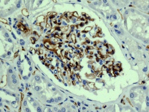
Immunohistochemical analysis of paraffin embedded Human kidney tissue labelling Integrin alpha 5 with ab150361 antibody at a dilution of 1/100.
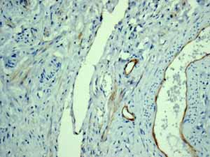
Immunohistochemical analysis of paraffin embedded Human Vessels tissue using ab150361 showing +ve staining.
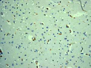
Immunohistochemical analysis of paraffin embedded Human Brain vessels tissue using ab150361 showing +ve staining.
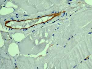
Immunohistochemical analysis of paraffin embedded Human Skeletal muscle vessel tissue using ab150361 showing +ve staining.
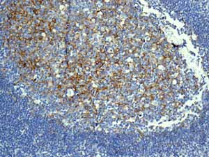
Immunohistochemical analysis of paraffin embedded Normal Human Tonsil tissue using ab150361 showing +ve staining.
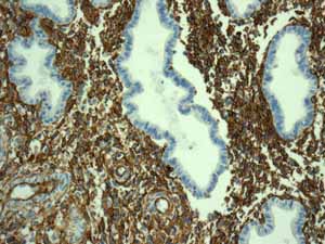
Immunohistochemical analysis of paraffin embedded Normal Human Uterus tissue using ab150361 showing +ve staining.
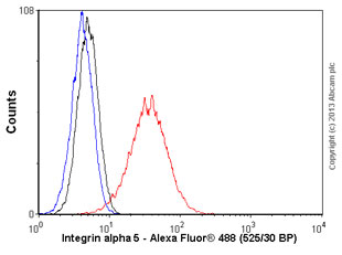
Overlay histogram showing HeLa cells stained with ab150361 (red line). The cells were fixed with 4% paraformaldehyde (10 min) and then permeabilized with 0.1% PBS-Tween for 20 min. The cells were then incubated in 1x PBS / 10% normal goat serum / 0.3M glycine to block non-specific protein-protein interactions followed by the antibody (ab150361, 1/1000 dilution) for 30 min at 22°C. The secondary antibody used was goat anti-rabbit Alexa Fluor 488 IgG (H&L) (ab150077) at 1/2000 dilution for 30 min at 22°C. Isotype control antibody (black line) was rabbit IgG (monoclonal) (0.1μg/1x106 cells) used under the same conditions. Unlabelled sample (blue line) was also used as a control. Acquisition of >5,000 events were collected using a 20mW Argon ion laser (488nm) and 525/30 bandpass filter. This antibody gave a positive signal in HeLa cells fixed with 80% methanol (5 min)/permeabilized with 0.1% PBS-Tween for 20 min used under the same conditions.