Western blot analysis of extracts from various cell lines using Btk (D3H5) Rabbit mAb.

Immunohistochemical analysis of paraffin-embedded human colon carcinoma using Btk (D3H5) Rabbit mAb. Note staining of inflammatory cells.

Immunohistochemical analysis of paraffin-embedded human B-cell lymphoma using Btk (D3H5) Rabbit mAb.

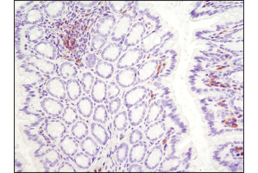
Immunohistochemical analysis of paraffin-embedded mouse colon using Btk (D3H5) Rabbit mAb.

Immunohistochemical analysis of paraffin-embedded human ovarian carcinoma using Btk (D3H5) Rabbit mAb. Note staining of inflammatory cells.

Immunohistochemical analysis of paraffin-embedded cell pellets, Ramos(left) or Jurkat (right), using Btk (D3H5) Rabbit mAb.

Human whole blood was fixed, lysed, and permeabilized as per the Cell Signaling Technology Flow Alternate Protocol and stained using Btk (D3H5) Rabbit mAb. Samples were co-stained using CD3-PE and CD19-APC to distinguish T and B cell subpopulations, respectively. B (red) and T (blue) cell population gates were applied to a histogram depicting the mean fluorescence intensity of Btk. Anti-rabbit IgG (H+L), F(ab') 2 Fragment (Alexa Fluor ® 488 Conjugate) #4412 was used as a secondary antibody.


