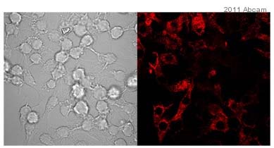
Anti-IL10 antibody
| Name | Anti-IL10 antibody |
|---|---|
| Supplier | Abcam |
| Catalog | ab9969 |
| Prices | $398.00 |
| Sizes | 100 µg |
| Host | Rabbit |
| Clonality | Polyclonal |
| Isotype | IgG |
| Applications | WB ELISA B/N ICC/IF ICC/IF |
| Species Reactivities | Mouse, Rat |
| Antigen | Recombinant fragment, corresponding to amino acids 19/178 of Rat IL10 (available as ab9970 |
| Description | Rabbit Polyclonal |
| Gene | Il10 |
| Conjugate | Unconjugated |
| Supplier Page | Shop |
Product images
Product References
Chronic antidepressant desipramine treatment increases open field-induced brain - Chronic antidepressant desipramine treatment increases open field-induced brain
Wrona D, Listowska M, Kubera M, Majkutewicz I, Glac W, Wojtyla-Kuchta B, Plucinska K, Grembecka B, Podlacha M. Brain Res Bull. 2013 Oct;99:117-31.
Effects of acupuncture on Th1, th2 cytokines in rats of implantation failure. - Effects of acupuncture on Th1, th2 cytokines in rats of implantation failure.
Gui J, Xiong F, Li J, Huang G. Evid Based Complement Alternat Med. 2012;2012:893023.
Acetate reduces microglia inflammatory signaling in vitro. - Acetate reduces microglia inflammatory signaling in vitro.
Soliman ML, Puig KL, Combs CK, Rosenberger TA. J Neurochem. 2012 Nov;123(4):555-67.
Impaired induction of IL-10 expression in the lung following hemorrhagic shock. - Impaired induction of IL-10 expression in the lung following hemorrhagic shock.
Khadaroo RG, Fan J, Powers KA, Fann B, Kapus A, Rotstein OD. Shock. 2004 Oct;22(4):333-9.


