![Anti-Id1 antibody [EPR7098] (ab134163) at 1/1000 dilution (unpurified) + HepG2 cell lysate at 10 µgSecondaryPeroxidase-conjugated goat anti-rabbit IgG (H+L) at 1/1000 dilution](http://www.bioprodhub.com/system/product_images/ab_products/2/sub_3/6272_ab134163-239287-ab134163upwb.jpg)
Anti-Id1 antibody [EPR7098] (ab134163) at 1/1000 dilution (unpurified) + HepG2 cell lysate at 10 µgSecondaryPeroxidase-conjugated goat anti-rabbit IgG (H+L) at 1/1000 dilution
![Anti-Id1 antibody [EPR7098] (ab134163) at 1/2000 dilution (purified) + HepG2 cell lysate at 10 µgSecondaryPeroxidase-conjugated goat anti-rabbit IgG (H+L) at 1/1000 dilution](http://www.bioprodhub.com/system/product_images/ab_products/2/sub_3/6273_ab134163-239288-ab134163pwb.jpg)
Anti-Id1 antibody [EPR7098] (ab134163) at 1/2000 dilution (purified) + HepG2 cell lysate at 10 µgSecondaryPeroxidase-conjugated goat anti-rabbit IgG (H+L) at 1/1000 dilution
![All lanes : Anti-Id1 antibody [EPR7098] (ab134163) at 1/1000 dilution (unpurified)Lane 1 : Mouse brain tissue lysateLane 2 : Rat brain tissue lysateLysates/proteins at 10 µg per lane.SecondaryPeroxidase-conjugated goat anti-rabbit IgG (H+L) at 1/1000 dilution](http://www.bioprodhub.com/system/product_images/ab_products/2/sub_3/6274_ab134163-239289-ab134163upwb2.jpg)
All lanes : Anti-Id1 antibody [EPR7098] (ab134163) at 1/1000 dilution (unpurified)Lane 1 : Mouse brain tissue lysateLane 2 : Rat brain tissue lysateLysates/proteins at 10 µg per lane.SecondaryPeroxidase-conjugated goat anti-rabbit IgG (H+L) at 1/1000 dilution
![All lanes : Anti-Id1 antibody [EPR7098] (ab134163) at 1/2000 dilution (purified)Lane 1 : Mouse brain tissue lysateLane 2 : Rat brain tissue lysateLysates/proteins at 10 µg per lane.SecondaryPeroxidase-conjugated goat anti-rabbit IgG (H+L) at 1/1000 dilution](http://www.bioprodhub.com/system/product_images/ab_products/2/sub_3/6275_ab134163-239290-ab134163pwb2.jpg)
All lanes : Anti-Id1 antibody [EPR7098] (ab134163) at 1/2000 dilution (purified)Lane 1 : Mouse brain tissue lysateLane 2 : Rat brain tissue lysateLysates/proteins at 10 µg per lane.SecondaryPeroxidase-conjugated goat anti-rabbit IgG (H+L) at 1/1000 dilution
![All lanes : Anti-Id1 antibody [EPR7098] (ab134163) at 1/1000 dilution (unpurified)Lane 1 : HeLa cell lysatesLane 2 : 293T cell lysatesLane 3 : HepG2 cell lysatesLysates/proteins at 10 µg per lane.SecondaryHRP labelled goat anti-rabbit at 1/2000 dilution](http://www.bioprodhub.com/system/product_images/ab_products/2/sub_3/6276_Id1-Primary-antibodies-ab134163-1.jpg)
All lanes : Anti-Id1 antibody [EPR7098] (ab134163) at 1/1000 dilution (unpurified)Lane 1 : HeLa cell lysatesLane 2 : 293T cell lysatesLane 3 : HepG2 cell lysatesLysates/proteins at 10 µg per lane.SecondaryHRP labelled goat anti-rabbit at 1/2000 dilution
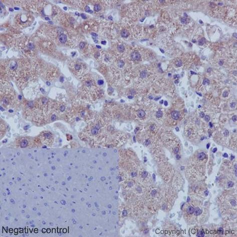
Immunohistochemistry (Formalin/PFA-fixed paraffin-embedded sections) analysis of human liver tissue labelling Id1 with unpurified ab134163 at 1/500. Heat mediated antigen retrieval was performed using Tris/EDTA buffer pH 9. A prediluted HRP-polymer conjugated anti-rabbit IgG was used as the secondary antibody. Negative control using PBS instead of primary antibody. Counterstained with Hematoxylin.
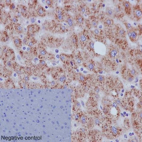
Immunohistochemistry (Formalin/PFA-fixed paraffin-embedded sections) analysis of human liver tissue labelling Id1 with purified ab134163 at 1/800. Heat mediated antigen retrieval was performed using Tris/EDTA buffer pH 9. A prediluted HRP-polymer conjugated anti-rabbit IgG was used as the secondary antibody. Negative control using PBS instead of primary antibody. Counterstained with Hematoxylin.
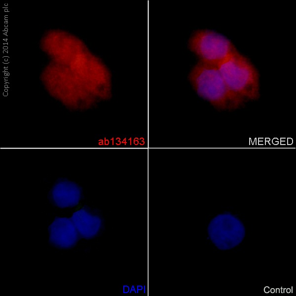
Immunocytochemistry/Immunofluorescence analysis of HepG2 cells labelling Id1 with unpurified ab134163 at 1/250. Cells were fixed with 4% paraformaldehyde. An Alexa Fluor® 555-conjugated goat anti-rabbit IgG (1/500) was used as the secondary antibody. DAPI (blue) was used as the nuclear counterstain.Control: primary antibody (1/250) and secondary antibody, ab150113, an Alexa Fluor® 488-conjugated goat anti-mouse IgG (1/500).
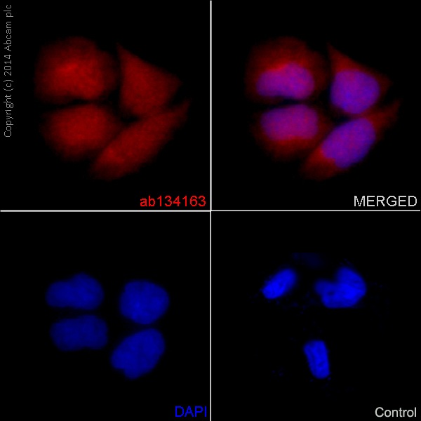
Immunocytochemistry/Immunofluorescence analysis of HepG2 cells labelling Id1 with purified ab134163 at 1/500. Cells were fixed with 4% paraformaldehyde. An Alexa Fluor® 555-conjugated goat anti-rabbit IgG (1/500) was used as the secondary antibody. DAPI (blue) was used as the nuclear counterstain.Control: primary antibody (1/250) and secondary antibody, ab150113, an Alexa Fluor® 488-conjugated goat anti-mouse IgG (1/500).
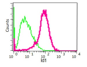
Flow cytometry analysis of HepG2 cells labelling Id1 with unpurified ab134163 at 1/90 (red). Cells were fixed with 2% paraformaldehyde. A FITC-conjugated goat anti-rabbit IgG (1/150) was used as the secondary antibody. Green - Isotype control, rabbit monoclonal IgG.
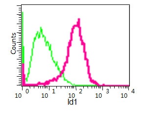
Flow cytometry analysis of HepG2 cells labelling Id1 with purified ab134163 at 1/160 (red). Cells were fixed with 2% paraformaldehyde. A FITC-conjugated goat anti-rabbit IgG (1/150) was used as the secondary antibody. Green - Isotype control, rabbit monoclonal IgG.