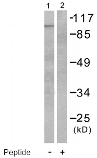
All lanes : Anti-Hsp105 antibody (ab53107) at 1/300 dilutionLane 1 : HeLa cell extractLane 2 : HeLa cell extract with immunising peptide

ab53107 at 1/50 dilution staining Hsp105 in human colon carcinoma by Immunohistochemistry, Paraffin embedded tissue, in the absence and presence of the immunising peptide.
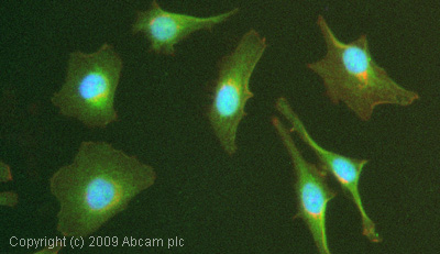
ICC/IF image of ab53107 stained HeLa cells. The cells were 4% PFA fixed (10 min) and then incubated in 1%BSA / 10% normal goat serum / 0.3M glycine in 0.1% PBS-Tween for 1h to permeabilise the cells and block non-specific protein-protein interactions. The cells were then incubated with the antibody (ab53107, 1æg/ml) overnight at +4øC. The secondary antibody (green)ÿwas Alexa Fluor© 488 goat anti-rabbit IgG (H+L) used at a 1/1000 dilution for 1h. Alexa Fluor© 594 WGA was used to label plasma membranes (red) at a 1/200 dilution for 1h. DAPI was used to stain the cell nuclei (blue) at a concentration of 1.43æM.