Anti-hnRNP U antibody - ChIP Grade
| Name | Anti-hnRNP U antibody - ChIP Grade |
|---|---|
| Supplier | Abcam |
| Catalog | ab20666 |
| Prices | $400.00 |
| Sizes | 100 µg |
| Host | Rabbit |
| Clonality | Polyclonal |
| Isotype | IgG |
| Applications | ICC/IF ICC/IF WB IHC-P ChIP |
| Species Reactivities | Mouse, Human, Rat |
| Antigen | Synthetic peptide conjugated to KLH derived from within residues 800 to the C-terminus of Human hnRNP U |
| Description | Rabbit Polyclonal |
| Gene | HNRNPU |
| Conjugate | Unconjugated |
| Supplier Page | Shop |
Product images
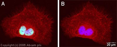 ICC/IF image of ab20666 stained human HeLa cells. The cells were methanol fixed (5 min) and incubated with the antibody (ab20666, 1µg/ml) for 1h at room temperature. The secondary antibody (green) was Alexa Fluor® 488 goat anti-rabbit IgG (H+L) used at a 1/1000 dilution for 1h. Image-iTTM FX Signal Enhancer was used as the primary blocking agent, 5% BSA (in TBS-T) was used for all other blocking steps. DAPI was used to stain the cell nuclei (blue). Alexa Fluor® 594 WGA was used to label plasma membranes (red). Panel A shows localisation of ab20666 to the nuclei, Panel B has the Alexa Fluor® 488 channel removed for comparison.
 Anti-hnRNP U antibody - ChIP Grade (ab20666) at 1 µg/ml + HeLa (Human epithelial carcinoma cell line) Whole Cell Lysate (ab27252) at 20 µgSecondaryGoat Anti-Rabbit IgG H&L (HRP) preadsorbed (ab7090) at 1/10000 dilutionPerformed under reducing conditions.
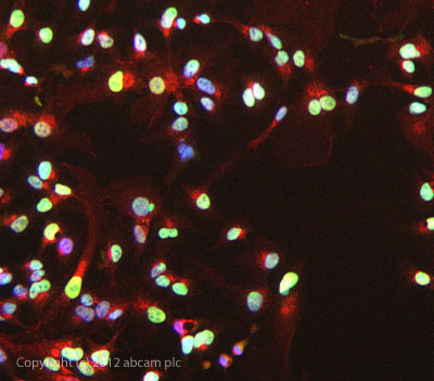
 Image courtesy of Human Protein Atlasab20666 staining hnRNP U in paraffin embedded human skin tissue. The samples were incubated with ab20666 (1/250 dilution) for 30 mins at room temperature. Antigen retrieval was performed by heat induction in citrate buffer pH 6. ab20666 was tested in a tissue microarray (TMA) containing a wide range of normal and cancer tissues as well as a cell microarray consisting of a range of commonly used, well characterised human cell lines.Further results for this antibody can be found at www.proteinatlas.org
 ab20666 stained HepG2 cells. The cells were 4% formaldehyde fixed (10 min) and then incubated in 1%BSA / 10% normal goat serum / 0.3M glycine in 0.1% PBS-Tween for 1h to permeabilise the cells and block non-specific protein-protein interactions. The cells were then incubated with the antibody ab20666 at 1µg/ml overnight at +4°C. The secondary antibody (green) was DyLight® 488 goat anti- rabbit (ab96899) IgG (H+L) used at a 1/1000 dilution for 1h. Alexa Fluor® 594 WGA was used to label plasma membranes (red) at a 1/200 dilution for 1h. DAPI was used to stain the cell nuclei (blue) at a concentration of 1.43µM.
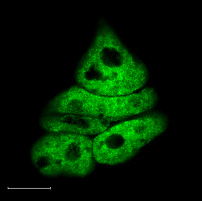 Immunofluorescence analysis of murine embryonic stem cells, staining hnRNP U with ab20666. Cells were fixed with paraformaldehyde, permeabilized with 0.25% Triton X-100 and blocked in 0.1% Triton X-100/10% FCS for 20 min. Samples were incubated with primary antibody (1/500) for 2 hours before incubating with an AlexaFluor®488-conjugated goat anti-rabbit IgG (1/500) for 1 hour.
Product References
Coordinate regulation of heterogeneous nuclear ribonucleoprotein dynamics by - Coordinate regulation of heterogeneous nuclear ribonucleoprotein dynamics by
Shao R, Wang X, Weijdegard B, Norstrom A, Fernandez-Rodriguez J, Brannstrom M, Billig H. Am J Physiol Endocrinol Metab. 2012 May 1;302(10):E1269-82. doi:
The expression profile of RNA-binding proteins in primary and metastatic - The expression profile of RNA-binding proteins in primary and metastatic
Hope NR, Murray GI. Hum Pathol. 2011 Mar;42(3):393-402.
SAF-A has a role in transcriptional regulation of Oct4 in ES cells through - SAF-A has a role in transcriptional regulation of Oct4 in ES cells through
Vizlin-Hodzic D, Johansson H, Ryme J, Simonsson T, Simonsson S. Cell Reprogram. 2011 Feb;13(1):13-27.
SAF-A forms a complex with BRG1 and both components are required for RNA - SAF-A forms a complex with BRG1 and both components are required for RNA
Vizlin-Hodzic D, Runnberg R, Ryme J, Simonsson S, Simonsson T. PLoS One. 2011;6(12):e28049.