Anti-hnRNP K antibody - ChIP Grade
| Name | Anti-hnRNP K antibody - ChIP Grade |
|---|---|
| Supplier | Abcam |
| Catalog | ab70492 |
| Prices | $388.00 |
| Sizes | 100 µl |
| Host | Rabbit |
| Clonality | Polyclonal |
| Isotype | IgG |
| Applications | WB IP IHC-P ICC/IF ICC/IF ChIP ChIP |
| Species Reactivities | Mouse, Human, Rat, Guinea Pig, Bovine, Monkey, Ape, Orangutan, Platypus, Elephant |
| Antigen | Synthetic peptide corresponding to a sequence from the C-terminus of isoform a of human hnRNP K |
| Description | Rabbit Polyclonal |
| Gene | HNRNPK |
| Conjugate | Unconjugated |
| Supplier Page | Shop |
Product images
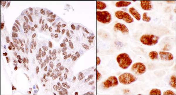 Immunohistochemistry (Formalin/PFA-fixed paraffin-embedded sections) analysis of human ovarian carcinoma (left) and mouse squamous cell carcinoma (right) tissues labelling hnRNP K with ab70492 at 1/1000 (0.2µg/ml). Detection: DAB.
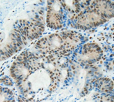 IHC image of hnRNP K staining in Human Colon Adenocarcinoma formalin fixed paraffin embedded tissue section, performed on a Leica Bond™ system using the standard protocol F. The section was pre-treated using heat mediated antigen retrieval with sodium citrate buffer (pH6, epitope retrieval solution 1) for 20 mins. The section was then incubated with ab70492, 0.5 µg/ml, for 15 mins at room temperature and detected using an HRP conjugated compact polymer system. DAB was used as the chromogen. The section was then counterstained with haematoxylin and mounted with DPX. For other IHC staining systems (automated and non-automated) customers should optimize variable parameters such as antigen retrieval conditions, primary antibody concentration and antibody incubation times.
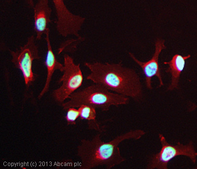 ICC/IF image of ab70492 stained HeLa cells. The cells were 100% methanol fixed (5 min) and then incubated in 1%BSA / 10% normal goat serum / 0.3M glycine in 0.1% PBS-Tween for 1h to permeabilise the cells and block non-specific protein-protein interactions. The cells were then incubated with the antibody (ab70492, 1µg/ml) overnight at +4°C. The secondary antibody (green) was ab96899, DyLight® 488 goat anti-rabbit IgG (H+L) used at a 1/250 dilution for 1h. Alexa Fluor® 594 WGA was used to label plasma membranes (red) at a 1/200 dilution for 1h. DAPI was used to stain the cell nuclei (blue) at a concentration of 1.43µM.
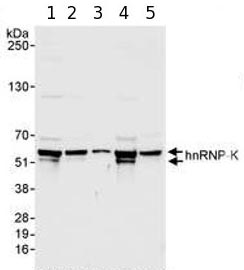 All lanes : Anti-hnRNP K antibody - ChIP Grade (ab70492) at 0.04 µg/mlLane 1 : HeLa whole cell lysate at 50 µgLane 2 : HeLa whole cell lysate at 15 µgLane 3 : HeLa whole cell lysate at 5 µgLane 4 : 293T whole cell lysate at 50 µgLane 5 : NIH3T3 whole cell lysate at 50 µg
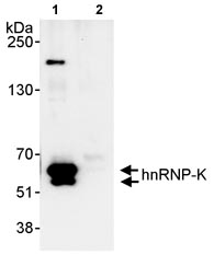 Detection of Human hnRNP K by Immunoprecipitation in Whole cell lysate from HeLa cells (1 mg/IP, 20% of IP loaded) using ab70492 at 3 µg/mg for IP and at 1.0 µg/ml for subsequent WB detection. Lane 2 represents IP IgG control.
Product References
hnRNP K coordinates transcriptional silencing by SETDB1 in embryonic stem cells. - hnRNP K coordinates transcriptional silencing by SETDB1 in embryonic stem cells.
Thompson PJ, Dulberg V, Moon KM, Foster LJ, Chen C, Karimi MM, Lorincz MC. PLoS Genet. 2015 Jan 22;11(1):e1004933.
A large intergenic noncoding RNA induced by p53 mediates global gene repression - A large intergenic noncoding RNA induced by p53 mediates global gene repression
Huarte M, Guttman M, Feldser D, Garber M, Koziol MJ, Kenzelmann-Broz D, Khalil AM, Zuk O, Amit I, Rabani M, Attardi LD, Regev A, Lander ES, Jacks T, Rinn JL. Cell. 2010 Aug 6;142(3):409-19.