
All lanes : Anti-Histone H4 (di methyl K79) antibody (ab2885) at 1/500 dilutionLane 1 : Calf Thymus histone prepLane 2 : Calf Thymus histone prep with Human Histone H3 (mono methyl K79) peptide (ab4555) at 0.1 µgLane 3 : Calf Thymus histone prep with Human Histone H3 (di methyl K79) peptide (ab4556) at 0.1 µgLane 4 : Calf Thymus histone prep with Human Histone H3 (tri methyl K79) peptide (ab4557) at 0.1 µgLane 5 : Calf Thymus histone prep with Human Histone H4 (di methyl K79) peptide (ab4560) at 0.1 µgLane 6 : Calf Thymus histone prep with Human Histone H3 (di methyl K27) peptide (ab1781) at 0.1 µgLysates/proteins at 1 µg per lane.SecondaryGoat Anti-Rabbit IgG H&L (HRP) (ab6721) at 1/2000 dilutionRabbit polyclonal to Histone H4 Di Methyl K79 at 1/500 on Calf Thymus histone prep (1 ug per lane).Lane 1: ab2885 1/500Lane 2: ab2885 1/500 H3 Mono Me K79 peptide (ab4555), 0.1 ugLane 3: ab2885 1/500 H3 Di Me K79 peptide (ab4556), 0.1 ugLane 4: ab2885 1/500 H3 Tri Me K79 peptide (ab4557), 0.1

SKN cells were stained with ab2885 (green) at a dilution of 1/200. The cells were fixed in paraformaldehyde for 10 minutes prior to incubation with ab2885. The DNA was stained with DAPI (blue). 100x magnification.

developed using the ECL techniquePerformed under reducing conditions.Exposure time : 2 minutesThis image is courtesy of an Abreview submitted by Dr Gerald Daviesab2885 at 1/2000 used in Western blot analysis of MCF7 cells (human breast cancer cells). Lane 1: MCF7 cell lysate control Lane 2: MCF7 cell lysate (cells treated with 50µm Troglitazone for 48 hours)Lane 3: MCF7 cells lysate (cells treated with 1µm Trichostatin for 48 hours)Troglitazone and trichostatin increase dimethyl H4 (K79) and act as anti-proliferatives in human breast cancer. The blot was stripped and reprobed with an alpha tubilin antibody to show loading equivalence.See Abreview
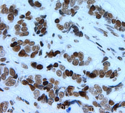
IHC image of Histone H4 (di methyl K79) staining in human breast carcinoma FFPE section, performed on a BondTM system using the standard protocol F. The section was pre-treated using heat mediated antigen retrieval with sodium citrate buffer (pH6, epitope retrieval solution 1) for 20 mins. The section was then incubated with ab2885, 5µg/ml, for 8 mins at room temperature and detected using an HRP conjugated compact polymer system. DAB was used as the chromogen. The section was then counterstained with haematoxylin and mounted with DPX.