![All lanes : Anti-Histone H4 (crotonyl K5) antibody [EPR17903] (ab177844) at 1/1000 dilutionLane 1 : NIH/3T3 (Mouse embyro fibroblast cells) cell lysateLane 2 : HeLa (Human epithelial cells from cervix adenocarcinoma) cell lysateLysates/proteins at 10 µg per lane.SecondaryGoat Anti-Rabbit IgG, (H+L), Peroxidase conjugated at 1/1000 dilution](http://www.bioprodhub.com/system/product_images/ab_products/2/sub_3/2281_ab177844-243068-AntiHistone-H4-crotonyl-K5--K8-antibody-EPR17903.jpg)
All lanes : Anti-Histone H4 (crotonyl K5) antibody [EPR17903] (ab177844) at 1/1000 dilutionLane 1 : NIH/3T3 (Mouse embyro fibroblast cells) cell lysateLane 2 : HeLa (Human epithelial cells from cervix adenocarcinoma) cell lysateLysates/proteins at 10 µg per lane.SecondaryGoat Anti-Rabbit IgG, (H+L), Peroxidase conjugated at 1/1000 dilution
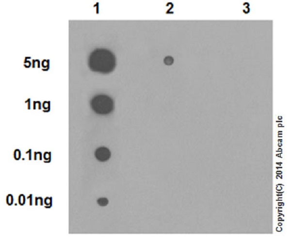
Dot blot analysis of crotonyl peptide (crotonyl K5) (Lane 1), crotonyl peptide (crotonyl K8) (Lane 2) and non- crotonyl peptide (Lane 3), labeled using ab177844 at 1/1000 dilution, followed by Goat Anti-Rabbit IgG, (H+L), Peroxidase conjugated secondary antibody at 1/1000 dilution.Blocking/Dilution buffer: 5% NFDM/TBST.
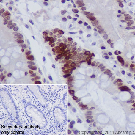
Immunohistochemical analysis of paraffin-embedded Human stomach tissue labeling Histone H4 (crotonyl K5 + K8) with ab177844 at 1/100 dilution, followed by Goat Anti-Rabbit IgG H&L (HRP) (ab97051) secondary antibody at 1/500 dilution. Nucleus staining on Human stomach tissue is observed. Counter stained with Hematoxylin.Secondary antibody only control: Used PBS instead of primary antibody, secondary antibody is Goat Anti-Rabbit IgG H&L (HRP) (ab97051) at 1/500 dilution.
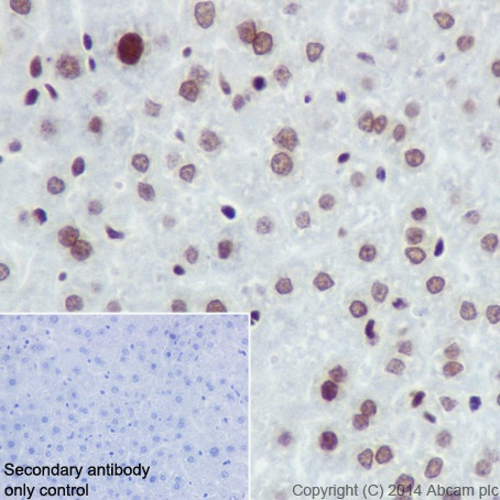
Immunohistochemical analysis of paraffin-embedded mouse liver tissue labeling Histone H4 (crotonyl K5 + K8) with ab177844 at 1/100 dilution, followed by Goat Anti-Rabbit IgG H&L (HRP) (ab97051) secondary antibody at 1/500 dilution. Nucleus staining on mouse liver tissue is observed. Counter stained with Hematoxylin.Secondary antibody only control: Used PBS instead of primary antibody, secondary antibody is Goat Anti-Rabbit IgG H&L (HRP) (ab97051) at 1/500 dilution.
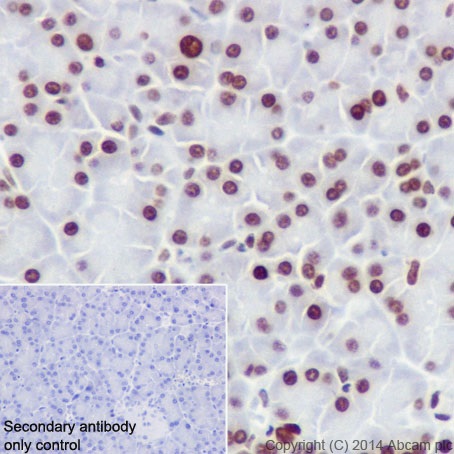
Immunohistochemical analysis of paraffin-embedded rat pancreas tissue labeling Histone H4 (crotonyl K5 + K8) with ab177844 at 1/100 dilution, followed by Goat Anti-Rabbit IgG H&L (HRP) (ab97051) secondary antibody at 1/500 dilution. Nucleus staining on rat pancreas tissue is observed. Counter stained with Hematoxylin.Secondary antibody only control: Used PBS instead of primary antibody, secondary antibody is Goat Anti-Rabbit IgG H&L (HRP) (ab97051) at 1/500 dilution.
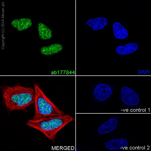
Immunofluorescent analysis of 4% paraformaldehyde-fixed, 0.1% Triton X-100 permeabilized HeLa (Human epithelial cells from cervix adenocarcinoma) cells labeling Histone H4 (crotonyl K5 + K8) with ab177844 at 1/250 dilution, followed by Goat anti-rabbit IgG (Alexa Fluor® 488) (ab150077) secondary antibody at 1/500 dilution (green). Confocal image showing nuclear staining on HeLa cell line. The nuclear counter stain is DAPI (blue). Tubulin is detected with ab7291 (anti-Tubulin mouse mAb) at 1/1000 dilution and ab150120 (AlexaFluor®594 Goat anti-Mouse secondary) at 1/500 dilution (red).The negative controls are as follows:-ve control 1: ab177844 at 1/250 dilution followed by ab150120 (AlexaFluor®594 Goat anti-Mouse secondary) at 1/500 dilution.-ve control 2: ab7291 (anti-Tubulin mouse mAb) at 1/1000 dilution followed by ab150077 (Alexa Fluor®488 Goat Anti-Rabbit IgG H&L) at 1/500 dilution.