ab61251 at 1/50 - 1/100 dilution staining Histone H3 in human lung carcinoma by Immunohistochemistry, Paraffin-embedded tissue, in the absence or presence of the immunising peptide.
All lanes : Anti-Histone H3 antibody (ab61251) at 1/500 dilutionLane 1 : RAW264.7 cell extracts treated with TSA (400nM, hours)Lane 2 : RAW264.7 cell extracts treated with TSA (400nM, hours) with the immunising peptide
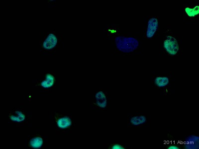
ab61251 staining Histone H3 in a human glioblastoma cell line by ICC/IF (Immunocytochemistry/immunofluorescence). Cells were fixed with paraformaldehyde, permeabilized with Triton X-100 (0.1%) in PBS and blocked with 0.5% BSA for 20 minutes at room temperature. Samples were incubated with primary antibody (1/50 in PBS + 0.5% BSA) for 16 hours at 4°C. An FITC-conjugated Goat anti-rabbit monoclonal (1/80) was used as the secondary antibody. Nuclei were counterstained with Hoechst (blue).See Abreview
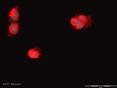
ab61251 staining Histone H3 in Rat choroid plexus cells by Immunocytochemistry/ Immunoflourescence. Cells were PFA-fixed and permeabilized in 0.1% Triton X-100 in PBS prior to blocking in 0.5% BSA in TBS-Tween for 20 minutes at room temperature. The primary antibody was diluted 1/50 in 0.5% BSA/PBS and incubated with the sample for 16 hours at 4°C. The secondary antibody was TRITC-conjugated Goat anti-Rabbit polyclonal, diluted 1/400. Nuclei were counterstained with Hoechst.See Abreview
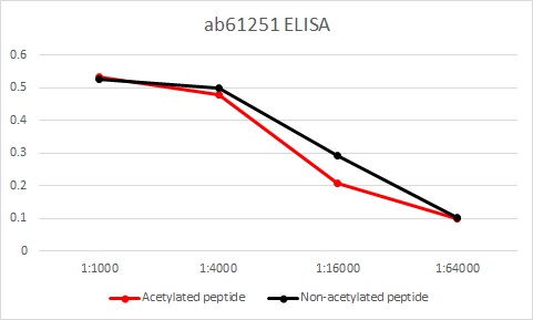
ELISA data for Anti-Histone H3 antibody, ab61251, which shows that the antibody at various dilutions detects both non-acetylated (black line) and acetylated (red line) histone H3.